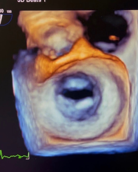
3D Transesophageal Echocardiography (3D TEE) is an advanced cardiac ultrasound technique that provides highly detailed, real-time three-dimensional images of the heart. Unlike conventional echocardiography, 3D TEE offers superior visualization of heart valves, chambers, and structural abnormalities, helping our doctor make more precise diagnoses and treatment decisions.
This minimally invasive imaging procedure is widely used for evaluating complex heart conditions, guiding interventional procedures, and planning cardiac surgeries with enhanced accuracy.
## What is 3D Transesophageal Echocardiography?
In a 3D TEE procedure, a specialized ultrasound probe is gently passed through the esophagus to obtain close and clear images of the heart. Since the esophagus lies directly behind the heart, this technique eliminates interference from the chest wall and lungs, resulting in exceptionally detailed cardiac imaging.
The addition of 3D technology allows us to assess cardiac structures from multiple angles in real time, improving diagnostic confidence and procedural precision.
## Benefits of 3D TEE
### Enhanced Visualization
3D imaging provides lifelike views of heart structures, especially heart valves and septal defects.
### Improved Diagnostic Accuracy
Helps detect complex cardiac abnormalities that may not be fully visible on standard 2D echocardiography.
### Better Surgical Planning
Supports cardiac surgeons and interventional cardiologists in planning minimally invasive and structural heart procedures.
### Real-Time Procedural Guidance
Frequently used during:
* Valve repair and replacement
* Mitral clip procedures
* ASD/VSD closure
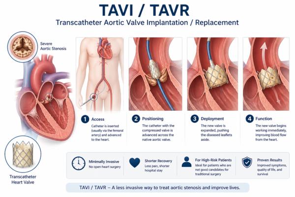
* TAVR procedures
* Left atrial appendage closure
### Minimally Invasive
The procedure is safe, relatively quick, and usually performed under mild sedation.
## Conditions Evaluated with 3D TEE
3D Transesophageal Echocardiography is commonly recommended for diagnosing and monitoring:
* Heart valve diseases
* Mitral valve prolapse
* Infective endocarditis
* Congenital heart defects
* Atrial septal defect (ASD)
* Ventricular septal defect (VSD)
* Blood clots inside the heart
* Structural heart abnormalities
* Aortic diseases
* Prosthetic valve assessment
## What to Expect During the Procedure
Before the procedure, patients are usually asked to fast for several hours. A mild sedative and local anesthetic spray are administered to ensure comfort.
During the examination:
1. A thin ultrasound probe is inserted through the mouth into the esophagus.
2. High-resolution 3D images of the heart are captured.
3. The procedure generally takes 20–45 minutes.
Most patients can return home the same day after a short observation period.
## Is 3D TEE Safe?
3D TEE is considered a safe and highly effective diagnostic procedure when performed by experienced cardiac specialists. Mild throat discomfort may occur temporarily after the test, but serious complications are rare.
## Why Choose Advanced 3D Cardiac Imaging?
Modern 3D echocardiography technology enables cardiologists to diagnose heart conditions earlier and with greater precision. This leads to:
* Faster treatment planning
* Improved surgical outcomes
* Better patient safety
* Enhanced cardiac care
## Book a 3D Transesophageal Echo Consultation
If you are experiencing symptoms such as chest discomfort, shortness of breath, palpitations, or have been advised to undergo advanced cardiac evaluation, 3D Transesophageal Echocardiography can provide detailed insights into your heart health.