An angiogram can map where a coronary artery narrows or closes, but the image does not answer every treatment question by itself. By the end, you will know how to read the main report terms, understand what the test can miss, and see how doctors connect an angiography finding to medication, a stent, bypass surgery, or no procedure.
Key takeaways
- Angiography shows where narrowing occurs, its length, and how many coronary arteries are affected.
- A 70% stenosis describes lumen narrowing, not the exact amount of plaque present.
- Use pressure or imaging tests when angiography alone cannot show whether narrowing limits blood flow.
- Treatment depends on symptoms, artery anatomy, blood flow, and overall health—not the percentage alone.
What the angiogram shows inside the coronary arteries
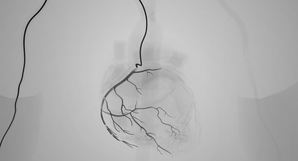
A coronary angiogram maps the contrast-filled inner channel, or lumen, of each coronary artery. It can show the artery’s diameter, route, branches, the number of affected vessels, and the stenosis location and length. These images also identify a coronary artery narrowing, a complete coronary artery blockage, or a severely narrowed segment.
| Finding on the image | Anatomical detail it can show | Important limitation |
|---|---|---|
| Narrowed segment | Location, apparent severity, length and vessel diameter | A single 2-D view can misjudge severity when arteries overlap, shorten, or contain heavy calcium |
| Complete occlusion | The blocked segment and sometimes collateral vessels feeding the artery beyond it | The image cannot establish when the blockage occurred, and collaterals may not provide normal flow |
| Abnormal vessel | Dissection, aneurysm, fistula, or an unusual coronary origin | Intravascular ultrasound, optical coherence tomography, or CT angiography may be needed for definition |
Doctors use multiple projections because one angle cannot display the coronary artery’s full three-dimensional shape. A visually estimated 70% narrowing is not automatically dangerous or a reason for a stent; an intermediate lesion may or may not restrict blood flow enough to cause ischemia.
The angiogram can also identify the artery related to a previous heart attack and show whether it remains occluded. It does not measure how much heart muscle remains viable or how strongly the ventricle contracts. Echo, cardiac MRI, or nuclear imaging answers those questions.
How to read the main terms in an angiography report
A 70% narrowing is not an automatic instruction to place a stent. The percentage is a visual estimate of lumen reduction against a nearby normal segment; it does not prove that blood flow is restricted enough to cause ischemia or symptoms.
1. Stenosis means narrowing inside an artery. A lesion is the abnormal area being described, which can include plaque, calcium, thrombus or a complete blockage. “Proximal” means closer to the artery’s origin; “distal” means farther along it. Reports may name the left main, LAD, LCx or RCA to identify the vessel and its segment.
2. TIMI flow describes contrast movement beyond the lesion: grade 0 means no flow, grade 1 means contrast enters but does not fully pass through, grade 2 means delayed flow, and grade 3 means normal flow. A severe-looking stenosis with preserved flow is not interpreted like an acute blockage with TIMI 0 flow.
3. Chronic total occlusion means a coronary artery has been completely blocked for at least three months. The image confirms the blockage but cannot reliably date it. Collateral vessels may supply the downstream heart muscle, although they do not guarantee normal circulation.
Doctors therefore combine the image with symptoms, ECG findings, heart function, lesion location and length, calcification, vessel size, and physiological testing when needed. A narrowing can be incidental, while a less dramatic lesion in the left main or another critical location can carry greater consequences.
What angiography cannot prove, and when another test is needed
An angiogram is a picture of the contrast-filled artery, not a complete measure of heart health. It may show an infarct-related artery that is blocked or severely narrowed, but it cannot prove how much heart muscle remains viable or how well the ventricle contracts.
A normal result also cannot exclude microvascular disease or coronary spasm, which can cause ischemic chest pain without a fixed blockage.
Use the additional test that answers the missing question:
- ECG: detects electrical changes from acute or previous myocardial injury, but a normal tracing does not exclude coronary disease.
- Troponin: identifies heart-muscle injury and helps diagnose acute myocardial infarction; it does not locate the blockage by itself.
- Echocardiography: measures ventricular contraction, valve function and regional wall-motion loss, including damage from a previous infarction.
- Stress testing: shows whether exertion or medication triggers ischemia; it helps connect a narrowing with symptoms.
- CT coronary angiography: provides noninvasive artery images and coronary calcium assessment, unlike invasive angiography, which is used for detailed lumen anatomy.
- IVUS: images the artery wall, plaque burden and stent expansion beyond the contrast-filled channel.
- OCT: supplies higher-resolution views of plaque disruption, thrombus and stent-related problems.
For an intermediate narrowing, fractional flow reserve of 0.80 or less, or an instantaneous wave-free ratio of 0.89 or less, supports flow limitation. A visible stenosis is not automatically the cause of symptoms; history and these additional findings must fit.
How doctors turn an angiography finding into a treatment decision
An angiogram does not automatically lead to a stent. Doctors match the narrowing to your symptoms, evidence of reduced blood flow, artery anatomy, heart function, medical risk and treatment preferences.
1. Medication and monitoring fit when the narrowing is mild, symptoms are controlled, or testing shows that blood flow is adequate. Treatment can include antiplatelet therapy, cholesterol-lowering medicine, blood-pressure control, diabetes management and anti-anginal drugs. “No procedure” means active risk reduction and follow-up, not ignoring the finding.
2. An intermediate narrowing needs a functional check before intervention. During angiography, FFR (fractional flow reserve) or iFR (instantaneous wave-free ratio) measures whether the lesion restricts blood flow; common thresholds are FFR 0.80 or lower and iFR 0.89 or lower. A 70% visual narrowing is not, by itself, a universal stent trigger.
3. A stent, or percutaneous coronary intervention, is considered when a lesion is flow-limiting, symptoms or ischemia persist, and the narrowing can be treated safely with a catheter. A short, accessible blockage is different from diffuse disease or a heavily calcified, tortuous segment, where stenting is more difficult.
4. Bypass surgery is considered for left-main disease, complex multivessel disease, difficult bifurcations or vessel origins, and anatomy where several stents would provide a less durable or less complete result. The heart team weighs surgical risk, heart function and overall disease burden.
Dr. Rajesh Matta can help connect the angiogram, physiological testing and symptoms before recommending an intervention. A narrowing in one artery is not automatically the cause of chest pain; spasm, microvascular disease or non-cardiac illness can produce similar symptoms.
Related service
Coronary Angiography & Angioplasty Coronary Angiography & Angioplasty At Anahat Advanced and Comprehensive Cardiac Clinic, Dr. Rajesh Matta provides advanced care for heart blockages through Coronary Angiography and... View service → |
What to expect before, during, and after coronary angiography
Ask exactly how to prepare, because fasting instructions and medication changes depend on your appointment time and health conditions.
1. Before the procedure, confirm when to stop food and clear liquids. Tell the team about diabetes, kidney disease, bleeding problems, pregnancy possibility, allergies to iodinated contrast, and every medicine you take, especially insulin, anticoagulants, and antiplatelet drugs. Do not stop a prescription unless they instruct you to.
2. During angiography, a local anaesthetic numbs the access site while you remain awake. The catheter usually enters through the radial artery at the wrist or the femoral artery in the groin.
You may feel pressure during insertion, brief warmth or flushing when contrast enters, and occasional palpitations; report chest pain, breathlessness, severe discomfort, or a new reaction immediately.
3. Ask how long observation will last and which access-site checks you will need. After radial access, a compression band remains around the wrist; after femoral access, you may need to lie flat while the puncture site is monitored for bleeding or swelling.
4. Before discharge, ask who will drive you home, when you may shower, and how long to avoid lifting, strenuous exercise, or bending at the hip. Keep the dressing clean, inspect the site, and seek urgent help for persistent bleeding, a growing lump, a cold or numb hand or leg, fever, or worsening chest symptoms.
Frequently asked questions
What does a coronary angiogram show?
It shows the contrast-filled lumen, route, branches, narrowing, complete blockages, affected vessels, and stenosis length.
What do stenosis and occlusion mean in an angiography report?
Stenosis means narrowing of an artery. Occlusion means the artery is completely blocked at the reported segment.
Can angiography prove that a narrowing causes reduced blood flow?
No. The image shows the lumen’s appearance, but fractional flow reserve or another physiological test may be needed to assess flow limitation.
How do doctors decide between medication, angioplasty, and bypass surgery?
They consider symptoms, blood-flow impact, lesion location and length, number of affected vessels, anatomy, and overall health.
What happens before and after coronary angiography?
You receive preparation instructions, contrast is injected through a wrist or groin catheter, and staff monitor you afterward for bleeding and kidney or allergic complications.