Calcium can make a coronary blockage rigid enough to stop a balloon from expanding or a stent from reaching its intended position. By the end, you will understand how doctors identify that mechanical problem, how rotablation prepares the artery, and when another calcium-modification method may be a better fit.
Key takeaways
- Rotablation creates a path for balloons and stents through rigid calcium.
- Doctors assess calcium thickness, length, vessel size and lesion anatomy before treatment.
- Rotablation differs from lithotripsy, orbital atherectomy and balloon angioplasty.
- Proper stent expansion and follow-up reduce the risk of recurring problems.
Why Calcium Makes a Coronary Blockage Hard to Treat
A heavily calcified coronary blockage contains calcium within the artery wall, sometimes as a hard, continuous ring around the vessel and sometimes as thick, irregular plates. This coronary artery calcification removes the wall’s flexibility, so the narrowed segment behaves more like a rigid pipe than soft plaque.
A standard balloon may expand at its ends while remaining pinched at the calcium, leaving a balloon-undilatable lesion. Increasing pressure does not reliably solve the problem; it can tear the artery or create a dissection while the calcium remains unmodified.
A stent needs a pathway wide enough for its delivery system, so a rigid bend or calcified shelf can create a stent-undeliverable lesion.
Even when the stent crosses, calcium can stop the metal frame from opening fully against the vessel wall. An underexpanded stent leaves a narrowed channel and increases the risk of tissue growing back through the stent or a clot forming inside it.
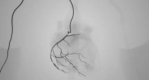
Angiography can underestimate this problem because it shows the vessel outline, not the calcium’s thickness or position. Intravascular ultrasound and optical coherence tomography show those details inside the artery.
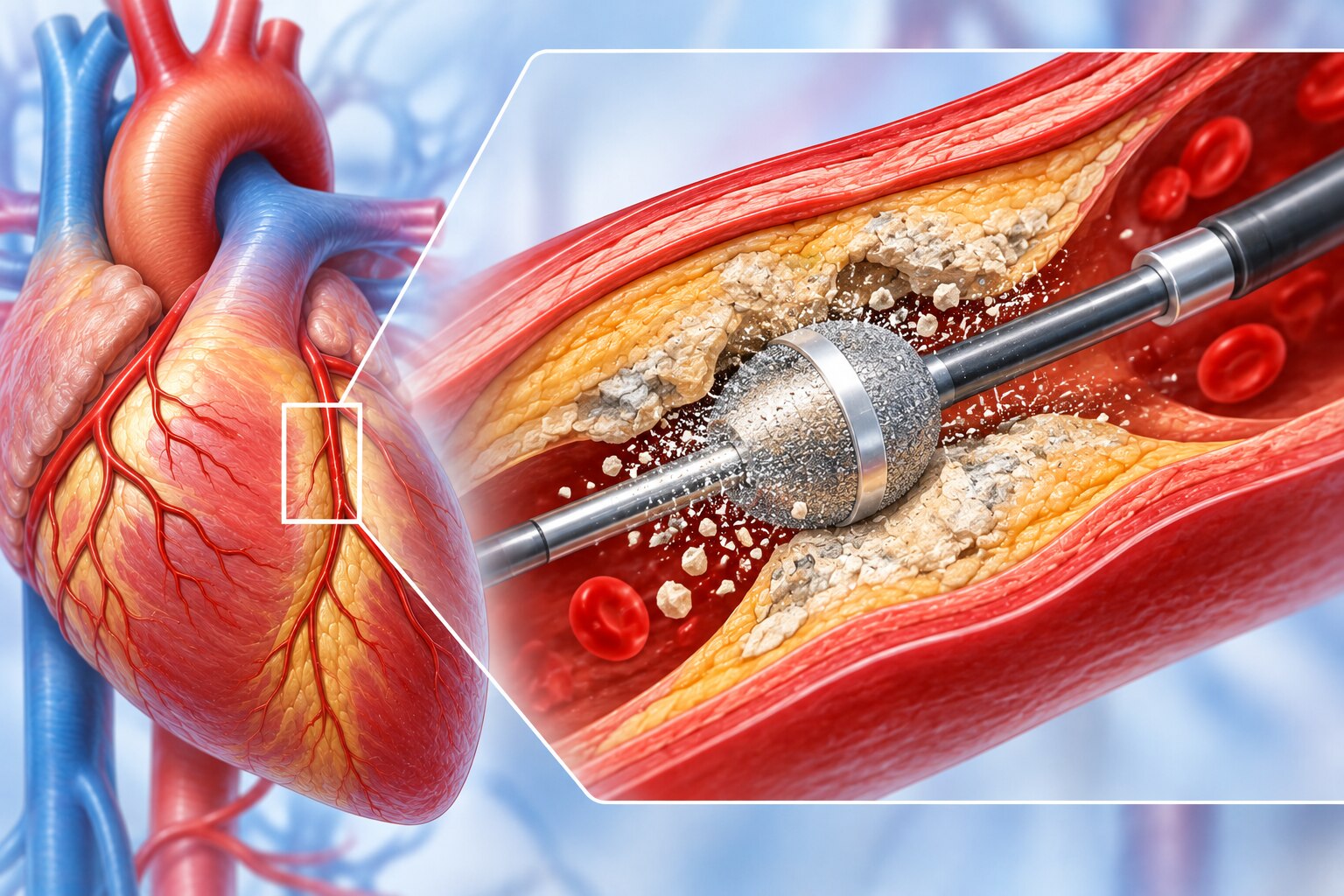
Rotablation addresses the mechanical obstacle by using a small diamond-coated burr to modify rigid calcium, creating a smoother path for balloon expansion and stent placement rather than removing the entire blockage.
How Doctors Decide Whether Rotablation Is Needed
Doctors consider rotablation for a wire-crossable lesion with severe calcium that a balloon cannot cross or expand, or when imaging predicts that calcium will prevent full stent expansion. Rotablation is not automatic: superficial versus deep calcium, vessel tortuosity, lesion length and available alternatives influence the choice.
Rotablation candidates commonly have:
- A tight, rigid narrowing where a balloon or stent stops before reaching the target.
- A balloon-undilatable lesion, even after an appropriately sized high-pressure balloon.
- A calcium pattern likely to leave an underexpanded stent, raising the risk of restenosis and stent thrombosis.
- A vessel route that permits burr delivery; if the guidewire cannot cross, another calcium-modification strategy is needed first.
An angiogram shows the artery’s outline, not the full calcium burden. Intravascular ultrasound uses a sound-based catheter to assess calcium arc, thickness, length and location, including deeper calcium that angiography can miss.
Optical coherence tomography provides sharper images of the calcium edge and superficial thickness, but it requires blood clearance and may not image beyond severe obstruction.
| Imaging method | What it shows | How it affects the decision |
|---|---|---|
| Angiography | Silhouette and apparent narrowing | Suggests calcium but can underestimate its depth and extent |
| Intravascular ultrasound | Arc, thickness, length, location and vessel dimensions | Identifies deep or extensive calcium and predicts expansion difficulty |
| Optical coherence tomography | High-resolution superficial calcium and lesion detail | Defines thin calcium deposits and stent-expansion targets when the catheter can cross |
If imaging shows limited calcium, balloons or lithotripsy may suffice; extensive, superficial, rigid calcium supports rotablation for lesion preparation.
What Happens During a Rotablation Procedure
Rotablation usually prepares a calcified lesion rather than removes the entire blockage. A catheter-mounted, diamond-coated burr spins at high speed and uses differential cutting: rigid calcium is abraded, while more elastic artery tissue deflects away. The fine particles usually pass through the microcirculation, but they can still trigger blood-flow problems.
1. The team obtains arterial access, usually through the radial or femoral artery, and guides a catheter to the coronary opening. Contrast images define the working path while the patient receives appropriate anticoagulation.
2. A specialised guidewire is advanced across the lesion. Guidewire passage can be difficult when calcium has narrowed or sharply angulated the artery; the burr catheter cannot reach the blockage until this wire is securely positioned.
3. The operator advances a small burr in short, controlled runs while rotating it, allowing blood flow and the burr system to cool between passes. This creates a smoother channel and modifies the calcium enough for balloon expansion.
4. A balloon is then inflated, followed by delivery of a drug-eluting stent when indicated. Further balloon post-dilation may be needed; intravascular imaging can confirm that calcium has not left the stent underexpanded.
Procedure-specific risks include coronary dissection or perforation, spasm, burr entrapment, distal embolization, slow or no-reflow, and access-site bleeding. Transient bradycardia or heart block deserves particular attention during right-coronary or dominant left-circumflex treatment. Contrast exposure and a longer procedure also matter for kidney risk, while stent implantation requires an antiplatelet plan.
Rotablation Versus Balloons, Orbital Atherectomy and Lithotripsy
The choice depends on whether the lesion can be crossed, whether a balloon can expand it, and where the calcium sits in the vessel wall.
| Option | What it does | When it fits |
|---|---|---|
| High-pressure balloon | Uses inflation force to crack resistant plaque | A balloon crosses and the calcium is limited or moderately rigid; excessive pressure risks dissection or perforation |
| Cutting balloon | Small blades make controlled incisions in plaque | A short, focal, balloon-crossable lesion needs a predictable opening |
| Scoring balloon | External wires or elements focus force along scoring lines | A resistant lesion needs controlled cracking with less uncontrolled balloon slippage |
| Rotablation | A diamond-coated burr modifies superficial calcium | The lesion is balloon-undilatable or stent-undeliverable, or imaging predicts severe stent underexpansion |
| Orbital atherectomy | An eccentrically rotating crown sands superficial calcium | A wire-crossable, heavily calcified lesion needs plaque modification across a suitable vessel size |
| Intravascular lithotripsy | Shock waves fracture calcium through a balloon | Deep, circumferential calcium is present and the balloon can cross and contact the vessel wall |
The anatomy can favour a combination: rotablation or orbital atherectomy to create passage, followed by a balloon; lithotripsy can then fracture deeper calcium before stenting. Doctors avoid atherectomy in an acutely thrombotic lesion, untreated dissection, or when the wire is not in the true lumen.
Dr. Rajesh Matta’s angioplasty planning should include intravascular imaging when angiography cannot show calcium depth or predict stent expansion. The burr passing is not the endpoint: balloon preparation, a drug-eluting stent, post-dilation and imaging may still be necessary.
Related service
Coronary Angiography & Angioplasty Coronary Angiography & Angioplasty At Anahat Advanced and Comprehensive Cardiac Clinic, Dr. Rajesh Matta provides advanced care for heart blockages through Coronary Angiography and... View service → |
Why Stent Expansion and Follow-Up Determine the Result
Passing the burr through the blockage is not the endpoint. The usual next step is balloon preparation followed by implantation of a drug-eluting stent, with post-dilation when needed to achieve full stent expansion and stent apposition against the artery wall.
An underexpanded or poorly apposed stent leaves gaps where tissue can grow or a clot can form.
Angiography can make the result look satisfactory while intravascular ultrasound or optical coherence tomography shows residual calcium, incomplete expansion, malapposition, inadequate lesion coverage or a dissection that needs treatment. Ask whether imaging was used to confirm the final result, especially when the artery was heavily calcified.
Discuss these issues before discharge and during follow-up:
| Issue | What to ask your cardiology team | Why it matters |
|---|---|---|
| Kidney function | How much iodinated contrast was used, and do I need hydration or a creatinine check? | Chronic kidney disease increases contrast-related kidney injury risk. |
| Bleeding | What is my bleeding risk, and which symptoms require urgent help? | Catheter access and antiplatelet treatment can cause or worsen bleeding. |
| Medication | When do I start and stop each antiplatelet drug, and what happens if I miss a dose? | Stopping prescribed therapy early raises stent-thrombosis risk. |
| Imaging | Was stent expansion and apposition adequate, and is repeat imaging needed? | A good angiographic picture alone cannot exclude mechanical failure. |
Continue prescribed cholesterol, blood-pressure, diabetes and smoking-cessation measures; the stent fixes one lesion, not the disease process.
Frequently asked questions
Why does calcium make a coronary blockage difficult to treat?
Calcium makes the artery wall rigid, so balloons may not expand the blockage and stents may fail to open fully.
How do doctors decide whether rotablation is needed?
Doctors assess the blockage using angiography and, when needed, intravascular imaging to measure calcium and plan treatment.
What happens during a rotablation procedure?
A catheter carries a tiny rotating burr across the calcified segment, where it modifies calcium before balloon treatment and stent placement.
How does rotablation compare with balloons, orbital atherectomy and lithotripsy?
Rotablation mechanically modifies calcium; balloons dilate the vessel, orbital atherectomy sands calcium with an eccentrically rotating crown, and lithotripsy uses sonic pressure waves.
Why do stent expansion and follow-up determine the result?
A fully expanded, well-positioned stent supports blood flow better, while follow-up checks symptoms, medicines and possible recurrent narrowing.