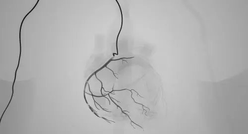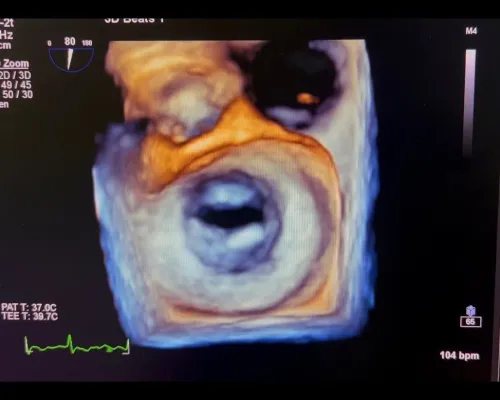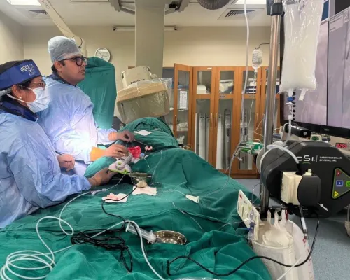

A narrowed coronary artery is not automatically the cause of chest pain, and a severe-looking blockage is not always best treated with a stent. By the end, you will understand which anatomical features make intervention difficult, which tests establish whether blood flow is truly restricted, and how doctors compare complex PCI with medication or bypass surgery.
Key takeaways
- Complexity depends on anatomy, access and treatment difficulty—not narrowing percentage alone.
- Pressure-wire testing can show whether a blockage is reducing blood flow.
- Ask how complex PCI compares with medication, standard PCI and bypass surgery.
- Confirm the expected benefits, risks, alternatives and recovery before proceeding.
Which coronary blockages are considered complex, and what prompts further assessment?
A blockage can be complex even when it is not the tightest narrowing on an angiogram. Complexity depends on the artery’s anatomy, how the lesion can be reached, and what treatment is required—not on the narrowing percentage alone.
- A chronic total occlusion is a completely blocked coronary artery present for at least 3 months.
- Heavy coronary calcium can prevent a balloon or stent from expanding properly.
- Severe tortuosity, very small arteries, long or diffuse disease, and multiple lesions make equipment delivery and treatment less reliable.
- Bifurcation disease affects a main vessel and its side branch; left-main disease threatens blood flow to a large amount of heart muscle.
- Vein-graft disease, previously stented segments, and arteries that are difficult to reach can require specialised planning and equipment.
| Feature | Why it raises complexity |
|---|---|
| Chronic total occlusion | The wire must cross an established complete blockage |
| Heavy calcium | The lesion may resist balloon expansion and stent delivery |
| Bifurcation or left-main disease | Treating one branch can compromise another or a large territory |
| Vein-graft disease or a previously stented segment | Old graft or stent structure can limit safe, reliable treatment |
| Very small or severely tortuous artery | Devices may not reach, cross, or open the target safely |
Further assessment is prompted by persistent exertional angina, worsening symptoms, reduced exercise capacity, objective ischaemia, or a large area of threatened heart muscle. Escalation also makes sense when an earlier procedure could not cross, expand, or treat the blockage reliably.
How do doctors prove that a blockage is reducing blood flow?
An angiogram shows the artery’s outline, but it cannot always prove which narrowing is reducing blood flow. An intermediate-looking lesion may be the culprit, an innocent bystander, or one of several contributors to symptoms.
| Test | What it measures | Common interpretation |
|---|---|---|
| FFR | Pressure beyond the narrowing during increased blood flow | FFR 0.80 or lower supports flow-limiting disease |
| iFR | Pressure change across the narrowing during a resting cardiac phase | iFR 0.89 or lower supports flow-limiting disease |
| Stress testing | Inducible ischaemia during exercise or medication-induced stress | Shows whether reduced blood flow occurs, and how much heart muscle is affected |
Fractional flow reserve and instantaneous wave-free ratio use a pressure wire passed across the lesion during catheterisation. They answer a local question: does this specific narrowing create a meaningful pressure drop? Stress testing answers a broader question by showing inducible ischaemia in a coronary territory, including its extent.
IVUS and OCT add detail that two-dimensional angiography can hide. They measure vessel diameter, show how calcium is distributed, map plaque length, identify suitable stent landing zones, and expose dissections or incomplete stent expansion. IVUS penetrates deeper through calcium; OCT provides finer surface detail but requires a blood-clearing contrast flush.
Together with pressure results, these images help determine whether revascularization is likely to relieve symptoms or protect a substantial area of heart muscle.
What happens during a planned complex PCI?
A planned complex PCI procedure begins with coronary CT or detailed angiographic views to map calcium, vessel diameter, bends, branch points, grafts and the safest route. The team selects radial or femoral access, guide catheters, wires, microcatheters and guide-extension catheters; femoral access may provide stronger support for difficult equipment.
- A guidewire is advanced across the lesion. Microcatheters support wire exchange, while re-entry devices help regain the true vessel channel after a controlled subintimal passage.
- The lesion is prepared with balloon angioplasty. High-pressure balloons treat resistant narrowing; cutting or scoring balloons create controlled plaque fractures before stenting.
- Heavy calcium may require rotational atherectomy, orbital atherectomy or intravascular lithotripsy. These methods modify and fracture calcium so a balloon and stent can pass and expand; they do not simply extract plaque.
- The operator deploys a drug-eluting stent and post-dilates it to fit the vessel.
| CTO strategy | What it means | When it applies |
|---|---|---|
| Antegrade | Advances through the blockage from its near side | The channel can be crossed directly |
| Dissection-and-re-entry | Travels around the blockage, then re-enters the true lumen | Direct wiring fails |
| Retrograde | Approaches through collateral vessels or a bypass graft | The distal cap is easier to reach |
At a bifurcation, a provisional one-stent strategy protects the side branch in most suitable lesions; a planned two-stent technique suits selected complex branches with extensive disease or calcium. IVUS or OCT then checks stent sizing, expansion, edge dissection and incomplete apposition before accepting the result.
When is complex PCI preferable to medication, standard PCI or bypass surgery?
A complex PCI is preferable only when it offers better symptom relief or revascularization than medication alone, standard PCI or surgery. Controlled symptoms and testing without significant ischaemia support medication and risk-factor treatment rather than an invasive procedure.
| Option | Strength | When it fits |
|---|---|---|
| Medication alone | Avoids procedural risks | Symptoms are controlled and tests show no significant ischaemia |
| Standard PCI | Treats a focal blockage simply | The lesion is accessible, crossable and expandable without specialised techniques |
| Complex PCI | Reaches or prepares difficult anatomy | Stents can safely treat the important vessel and surgery is less suitable |
| CABG | Can provide more complete, durable revascularization | Left-main disease, three-vessel or diffuse disease, extensive bifurcations, or multivessel disease with diabetes |
A Heart Team makes the complex PCI versus CABG decision when practical. It weighs:
- The angiogram and, where needed, IVUS or OCT; vessel anatomy; expected completeness of revascularization; and whether the blockage supplies viable heart muscle.
- Diabetes, kidney disease, previous bypass grafts, left-ventricular function, frailty and surgical risk.
- Your ability to take antiplatelet medication and your preference after the risks, recovery and alternatives are explained.
Do not choose a complex stent procedure from a blockage percentage alone. The assessment requires the angiogram, medical history, complete medication list and test results; Dr. Rajesh Matta can use those details to frame the treatment discussion rather than assume that a difficult-looking lesion needs PCI.
Related service
Coronary Angiography & Angioplasty Coronary Angiography & Angioplasty At Anahat Advanced and Comprehensive Cardiac Clinic, Dr. Rajesh Matta provides advanced care for heart blockages through Coronary Angiography and... View service → |
When can complex intervention be the wrong choice, and what should you ask before proceeding?
PCI can be the wrong choice when bypass surgery would treat the anatomy more completely, the artery supplies little viable heart muscle, or testing shows no significant ischaemia. Active bleeding or an inability to take dual antiplatelet therapy also weighs strongly against stent placement because early interruption can cause acute stent thrombosis.
A chronic total occlusion (CTO) is not automatically worth opening. CTO PCI may require more contrast, radiation, procedure time and technical equipment, with no guaranteed survival benefit.
A staged PCI can limit contrast exposure, radiation and physiologic stress; this approach is particularly useful with kidney disease and contrast concerns, extensive multivessel disease or a difficult CTO. Staging does not mean the first procedure failed.
Mechanical circulatory support is reserved for selected patients with severely reduced left-ventricular function, a large myocardium at risk or little ability to tolerate temporary ischaemia. Devices introduce their own hazards, including bleeding, vascular injury and limb ischaemia.
Before proceeding, ask:
- What is the FFR or iFR result, and does it show that this blockage is limiting blood flow?
- Is IVUS or OCT needed to measure the vessel, calcium and stent expansion safely?
- Would CABG provide more complete treatment than PCI for this anatomy?
- How will kidney function, diabetes and prior grafts change the contrast, access and staging plan?
- How will bleeding risk and my ability to continue dual antiplatelet therapy affect the choice and timing?
Frequently asked questions
Which coronary blockages are considered complex?
Complex blockages involve challenging anatomy, difficult access or specialised treatment, including heavily calcified lesions, chronic total occlusions, branching lesions and long or tortuous narrowing.
How do doctors prove that a blockage is reducing blood flow?
Doctors can use pressure-wire measurements such as fractional flow reserve or instantaneous wave-free ratio, alongside symptoms, stress testing and coronary imaging.
What happens during a planned complex PCI?
The team guides catheters to the coronary artery, crosses or prepares the blockage, restores the vessel opening and places a stent when appropriate. Intravascular imaging and specialised devices may be used.
When is complex PCI preferable to medication, standard PCI or bypass surgery?
The choice depends on symptoms, blood-flow evidence, anatomy, heart function, other illnesses and procedural risk. A heart team compares complex PCI with medication, standard PCI and coronary artery bypass surgery.
When can complex intervention be the wrong choice?
It may be unsuitable when symptoms are not caused by the blockage, blood flow is adequate, procedural risk is excessive or bypass surgery offers a safer and more durable option.