A conventional echocardiogram can show important slices of the heart, but complex valve disease often depends on understanding the full three-dimensional shape and position of the abnormality. By the end, you will know how 3D echocardiography creates that view, when transthoracic or transesophageal imaging is used, and how to read the measurements and findings in context.
Key takeaways
- 3D echo reconstructs heart anatomy from ultrasound data captured across a heartbeat.
- 3D TEE provides closer valve views when chest-wall imaging cannot answer the question.
- Valve shape, motion, leaks and surrounding structures matter more than one measurement.
- 3D echo helps plan valve procedures and check results for complications.
How 3D echocardiography creates a volume of the heart
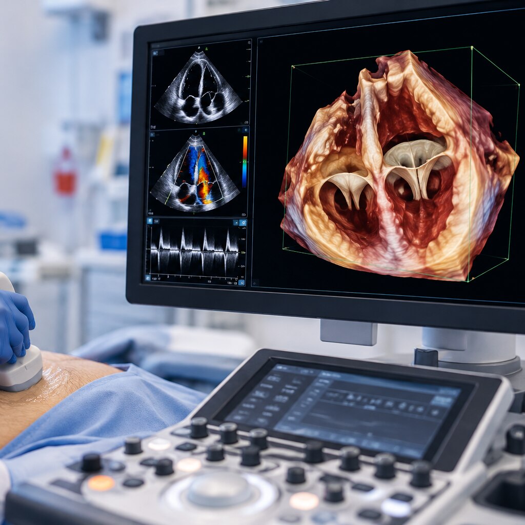
A standard ultrasound image is a thin, flat slice through the heart. 3D echocardiography uses a matrix-array probe to capture ultrasound echoes from multiple angles across a heartbeat, then reconstructs them as small volume elements called voxels. The result is a 3D echo dataset: a volume of cardiac anatomy rather than one fixed screenshot.
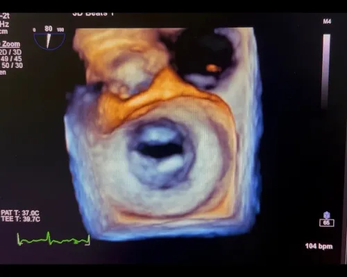
Three-dimensional cardiac imaging lets the clinician reorient that volume and inspect it in different planes. A valve can be viewed en face from the atrial or ventricular side, while its position relative to the annulus and nearby structures remains visible.
Volumetric echocardiography also supports chamber-volume measurements without estimating the chamber from a single diameter or assumed shape.
The volume does not replace 2D imaging or Doppler. Two-dimensional views capture rapid motion clearly, while Doppler measures blood-flow velocity and direction. A 3D TTE acquisition through the chest can lose detail when ribs, lung tissue or body habitus block the beam, and rotating a poor dataset cannot restore missing information.
The full volume must be reviewed, not just a favourable cropped image.
3D TTE or 3D TEE: which route provides the clearest view?
The clearest view depends on the question and the available acoustic window: 3D TEE usually shows valve anatomy more sharply, while 3D TTE is easier and safer to perform.
| Option | Image access | Practical trade-off |
|---|---|---|
| 3D TTE | The probe remains on the chest wall. It can measure chamber volumes and show valves during a routine, noninvasive examination. | Obesity, chronic lung disease, narrow rib spaces, dressings, and prosthetic material can block or distort the volume. A poor dataset cannot be fixed by rotating or cropping it. |
| 3D TEE | The probe sits in the oesophagus, close to the heart, so lungs and the chest wall interfere less. It provides detailed views of mitral, tricuspid, aortic, and prosthetic valves. | The test is semi-invasive. You must swallow the probe, and sedation is often used; anesthesia is required for some procedures. |
3D transesophageal echocardiography is preferred when leaflet tears, small perforations, commissures, device position, or procedural guidance demands a high-resolution view. 3D transthoracic echocardiography is the sensible first route for many outpatient assessments and serial chamber measurements.
Neither replaces everything else. Clinicians combine 3D TTE or 3D TEE with 2D imaging and Doppler, which capture rapid motion, blood-flow jets, velocity, and pressure-related measurements more effectively.
What 3D echo shows that a single flat slice can miss
A single 2D slice can show part of a valve while hiding the lesion’s position, shape, and relationship to nearby structures. A 3D dataset lets the clinician rotate and crop the heart, then view a valve en face from the atrial or ventricular side.
| Structure or measurement | What 2D may miss | What 3D makes easier to assess |
|---|---|---|
| Mitral valve | The full shape and movement of the mitral valve leaflets | Commissures, clefts, perforations, prolapse, flail segments, and leaflet number |
| Mitral annulus | One diameter from a single plane | The nonplanar annulus, annular dimensions, leaflet tethering, and coaptation |
| Regurgitant opening | An assumption that the leak is circular | The three-dimensional vena-contracta area, location, and irregular shape |
| Chambers and other valves | A shortened or incomplete view | Left atrium volume, left ventricular shape, tricuspid anatomy, aortic cusps, septal defects, and prosthetic-valve relationships |
This view is particularly useful when planning repair because it shows coaptation length, grasping zones, commissural location, and the predicted device relationship with the mitral orifice. It does not replace Doppler: flow velocity, transmitral gradient, and residual leakage still require haemodynamic assessment. Interpret the complete volume and multiplanar views, not one attractive screenshot.
Reading valve anatomy in mitral, aortic and tricuspid disease
Three-dimensional imaging clarifies valve disease by showing the valve en face and measuring an abnormal opening in its actual shape, rather than inferring anatomy from one flat slice. In mitral regurgitation, 3D colour Doppler can display the location and shape of the regurgitant orifice and calculate its vena contracta area.
An oval or crescent-shaped opening can therefore be assessed without assuming it is circular. Gain, Nyquist limit, spatial resolution, jet eccentricity and loading conditions still affect the measurement.
An en-face view can show:
- Mitral leaflet prolapse, flail segments, clefts or perforations, and their relationship to the annulus
- Aortic cusp number, commissures, raphe, opening pattern and aortic-root geometry
- Tricuspid leaflet position, commissures and coaptation gaps, including complex or prosthetic anatomy
For valve stenosis, 3D shows cusp restriction, calcification, commissural fusion and the three-dimensional orifice; Doppler velocity, pressure gradient and valve area determine haemodynamic severity. Heavy aortic calcium can create acoustic shadowing or blooming, hiding the true cusp edges and orifice.
A large prolapse or restricted leaflet is an anatomical finding, not a severity verdict: interpretation also uses flow, chamber response, symptoms, blood pressure, rhythm and loading conditions.
How 3D echo supports valve procedures and detects complications
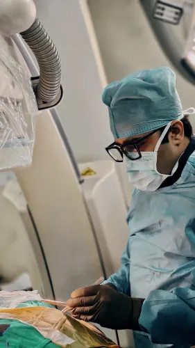
3D transesophageal echocardiography (3D TEE) guides structural valve procedures because it shows the valve en face and displays the device, leaflets and surrounding structures in one volume. It complements fluoroscopy, pressure measurements and Doppler rather than replacing them.
1. For TAVI planning, 3D echo assesses aortic cusp number, commissures, annular shape and aortic-root relationships. Heavy calcium can create shadowing and make the true orifice or cusp boundaries difficult to define, so CT remains important for annular sizing and access planning.
During implantation, 3D TEE helps confirm prosthesis position and detect paravalvular regurgitation, pericardial effusion or new interference with nearby structures.
2. Before a MitraClip procedure, 3D TEE maps the prolapse, flail segment or restricted leaflet and identifies the intended grasping zone. During deployment, it shows leaflet capture, device orientation, coaptation and the residual mitral opening; Doppler confirms residual regurgitation and transmitral gradient.
3. TriClip uses the same principles for tricuspid repair, but the large, irregular annulus and right-sided anatomy make spatial guidance especially valuable. Imaging checks leaflet capture and residual tricuspid regurgitation, while monitoring can reveal leaflet injury, device malposition, obstruction or pericardial fluid.
How to interpret a 3D echo report without relying on one number
A meaningful 3D echo interpretation matches anatomy, blood flow, heart response and symptoms rather than treating one abnormal number as the diagnosis.
1. Chamber volumes show how much blood the atria and ventricles hold. Enlarged chambers can indicate longstanding valve leakage or pressure overload, but the finding matters only alongside rhythm, body size, filling pressures and symptoms.
2. Ejection fraction describes the percentage of blood the left ventricle pumps out with each contraction. A preserved ejection fraction does not exclude serious valve disease; the ventricle can compensate while chamber volumes, strain or symptoms worsen.
3. Valve area estimates the opening available for blood flow. Pressure gradients show the force required to move blood through it, so both must be read with stroke volume, heart rate and blood pressure; low flow can produce a deceptively modest gradient.
4. Residual leakage after repair or replacement depends on the leak’s location, size, direction and effect on chamber volumes. For prosthetic valves, compare Doppler findings with valve type, position and prior studies rather than using an isolated gradient.
Dr. Rajesh Matta can help reconcile the 3D contours with Doppler data and clinical findings when automated measurements or image quality leave the report uncertain.
Related services
2D Echocardiography & Doppler Study 2D Echocardiography & Doppler Study At Anahat Advanced and Comprehensive Cardiac Clinic, 2D Echocardiography & Doppler Study is performed by Dr. View service → |
ECG (Electrocardiogram) At Anahat Advanced & Comprehensive Cardiac Clinic, ECG (Electrocardiogram) is performed by Dr Rajesh Matta to evaluate the heart’s electrical... View service → |
Frequently asked questions
How does 3D echocardiography create a volume of the heart?
A matrix-array probe captures ultrasound echoes from multiple angles across a heartbeat. Software reconstructs those data into a volume made of small elements called voxels.
Is 3D TTE or 3D TEE better for viewing heart valves?
3D TTE uses a probe on the chest and is non-invasive. 3D TEE places the probe in the oesophagus, closer to the heart, and often gives a clearer view when chest-wall images are limited or detailed valve planning is needed.
What can 3D echo show that a flat ultrasound slice can miss?
A 3D dataset can show valve shape, leaflet gaps, the direction and location of abnormal openings, and the relationship between valves and nearby structures.
How does 3D echo help with mitral, aortic and tricuspid valve disease?
It helps clinicians examine leaflet motion, valve opening, abnormal narrowing, leaks, prolapse, holes and surrounding anatomy in three dimensions.
How is 3D echo used during valve procedures?
It supports procedure planning, device positioning and assessment of the result. It also helps detect residual leaks, obstruction, perforation or other complications.
Related services
Structural Heart Procedures – TAVI, MitraClip & TriClip Structural Heart Procedures – TAVI, MitraClip & TriClip At Anahat Advanced & Comprehensive Cardiac Clinic, Dr. Rajesh Matta offers advanced structural heart interventions for patients with complex valve... View service → |
Heart Valve Disorder Treatment Heart Valve Disorder Treatment At Anahat Advanced and Comprehensive Cardiac Clinic, Dr. Rajesh Matta provides expert diagnosis and advanced treatment for heart valve disorders such... View service → |
Post-Covid Cardiac Complications Treatment Post-Covid Cardiac Complications Treatment At Anahat Advanced and Comprehensive Cardiac Clinic, Dr. Rajesh Matta offers specialized evaluation and treatment for post-Covid cardiac... View service → |
Cardiac Consultation & Evaluation Cardiac Consultation & Evaluation At Anahat Advanced and Comprehensive Cardiac Clinic, Dr. Rajesh Matta offers expert Cardiac Consultation & Evaluation for patients experiencing chest... View service → |