Calcium inside a coronary artery can make a balloon difficult to expand and leave a stent partly crushed or underexpanded. You will learn how doctors identify that problem, how orbital atherectomy modifies the plaque, what the procedure involves, and which risks and follow-up decisions deserve attention.
Key takeaways
- Calcium can stop a balloon from crossing or fully expanding a coronary blockage.
- An orbital crown sands and modifies calcium so a stent can expand more reliably.
- Doctors assess imaging, blockage severity, vessel size, and balloon response before atherectomy.
- Ask about alternatives, benefits, risks, recovery, and how success will be measured.
Why Calcium Makes Coronary Angioplasty Harder
Coronary artery calcification is hardened calcium within an atherosclerotic plaque, often forming a rigid ring or shelf inside the artery. In a calcified coronary blockage, balloon angioplasty may fail because the balloon cannot cross the lesion or cannot expand it enough to open the artery.
A stent may then fail to reach the target or seat properly.
Rigid calcium also resists the force needed to expand a stent. The result, called stent underexpansion, leaves a narrow channel and raises the risks of dissection, perforation, slow flow, restenosis and stent thrombosis.
1. Orbital atherectomy is usually preparation, not a cure. It modifies calcium so a balloon can expand and a drug-eluting stent can be delivered and opened more completely; it does not restore a normal artery or eliminate atherosclerosis by itself.
2. The operator may use it when a balloon or imaging catheter cannot cross, when the lesion will not expand, or when imaging predicts poor stent expansion. A technically successful result still needs final imaging or angiography to check for residual calcium and inadequate expansion.
3. After stenting, continue prescribed antiplatelet treatment. Cholesterol lowering, blood-pressure and diabetes control, smoking cessation and follow-up remain essential because the underlying disease continues beyond the treated segment.
How the Orbital Crown Modifies the Plaque
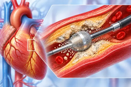
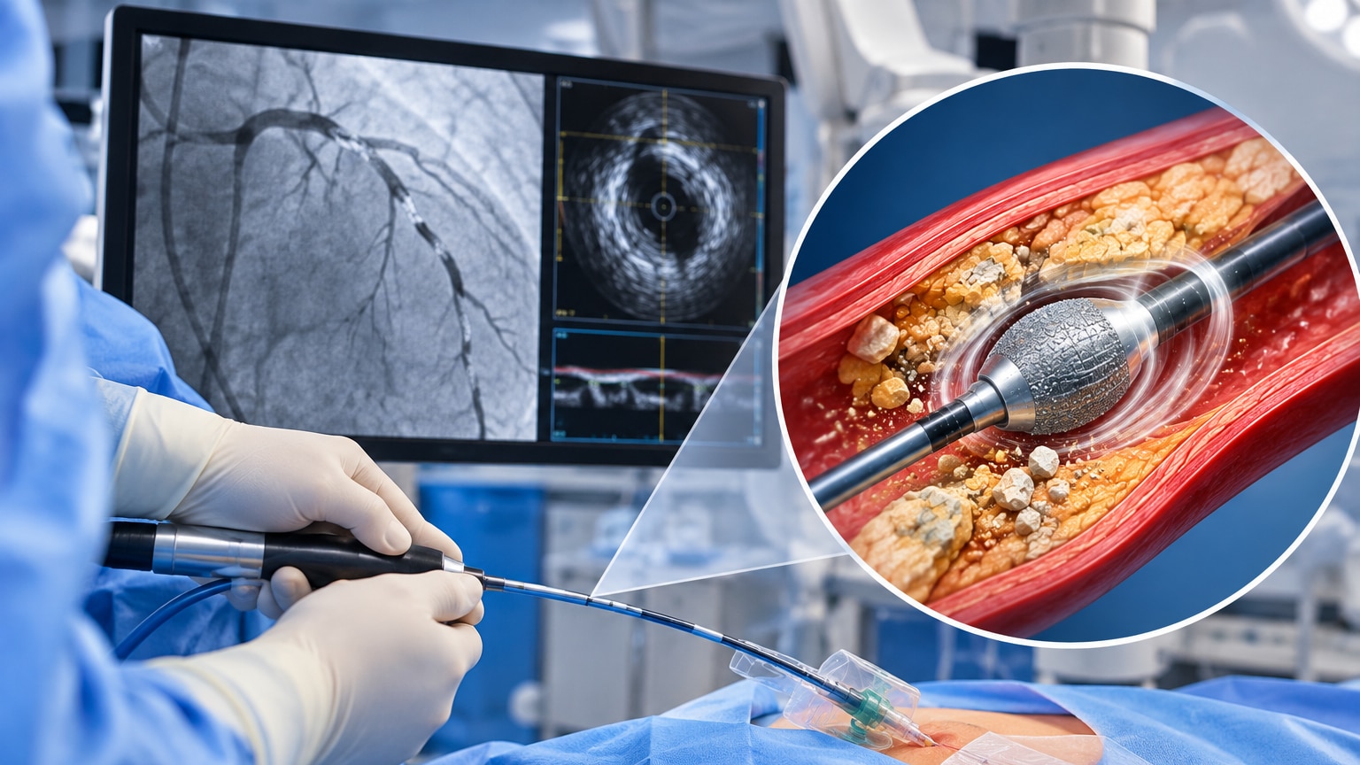
A specialized guidewire carries the orbital system through the narrowed coronary artery. Its diamond-coated crown is mounted eccentrically, so it rotates around the wire rather than spinning centrally inside the vessel.
Centrifugal force presses the crown against raised, harder calcium more than against softer plaque and vessel tissue. This selective action, called differential sanding, modifies the rigid surface instead of aggressively cutting the entire arterial wall.
The operator uses brief, controlled runs while continuously infusing saline. The crown is advanced and withdrawn rather than held motionless, reducing heat and deep vessel injury. Each run creates microscopic particulate debris and a more workable channel through the lesion.
A balloon then prepares the segment, followed usually by placement and expansion of a drug-eluting stent.
Orbital atherectomy and rotational atherectomy use different mechanisms:
| Approach | Device movement | Practical distinction |
|---|---|---|
| Orbital atherectomy | Eccentrically orbiting diamond-coated crown | Sands raised calcium while orbiting around the guidewire |
| Rotational atherectomy, or rotablation | Centrally rotating diamond-coated burr | Abrades plaque at the burr’s rotating tip |
The choice depends on lesion length and calcium pattern, vessel size, tortuosity, angulation, wire access, device support, and whether the equipment can cross and expand the lesion safely. A heavily calcified artery does not automatically require atherectomy; a balloon-based method may be enough when the lesion crosses and expands adequately.
How Doctors Decide Whether Atherectomy Is Needed
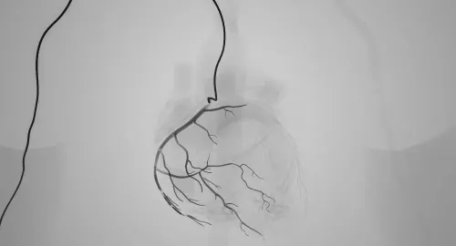
A difficult-looking angiogram does not prove that atherectomy is necessary. Angiography mainly shows radiopaque calcium shadows, and vessel overlap can hide calcium or make a lesion appear more severe than it is. It also cannot reliably show calcium thickness or predict whether a stent will expand.
IVUS calcium assessment shows the artery from inside, including calcium arc, thickness, length and vessel diameter. Optical coherence tomography provides higher-resolution images of the calcium surface and helps assess whether cracks or modification could permit expansion.
A frequently used IVUS marker is calcium extending beyond a calcium arc 270 degrees over at least 5 mm; thick, long and nearly circumferential calcium raises concern for balloon and stent underexpansion.
No single measurement automatically mandates atherectomy. The team also considers whether the device can cross and whether the lesion responds to low-pressure balloon inflation:
- If a small balloon crosses and expands the lesion adequately, balloon-based calcium modification or another non-atherectomy method may be enough.
- If the balloon or intravascular imaging catheter cannot cross, atherectomy becomes more persuasive.
- If the lesion will not expand despite balloon treatment, further calcium modification may be needed.
- If imaging predicts dangerous stent underexpansion, atherectomy can improve lesion preparation before stenting.
The goal is not to remove all plaque. It is to create enough compliance for the balloon and stent to cross, expand and contact the vessel wall safely.
What the Procedure Feels Like and How It Proceeds
Your experience varies with access site, anatomy, sedation and treatment needs, but the usual sequence is predictable. Follow fasting instructions and ask which medicines to take or hold; never stop antiplatelet or diabetes medicines without specific advice. You sign consent after discussing benefits, risks and alternatives.
1. A nurse places monitoring and an intravenous line. The cardiologist numbs the wrist or groin with local anaesthetic, then uses radial access or femoral access to enter the artery. You receive anticoagulation to reduce clot formation, and the team explains that contrast dye and X-ray radiation are involved.
2. Coronary angiography shows the blockage. A specialised guidewire is navigated across it during guidewire crossing, and IVUS or OCT provides intravascular images when the catheter can pass safely. Remain still, and report chest discomfort or breathing difficulty immediately.
3. If calcium requires modification, the operator advances the diamond-coated crown over the wire and makes short runs, advancing and withdrawing it rather than holding it still. Saline flows continuously. A low-pressure balloon then prepares the vessel; additional balloons, stents or another treatment can become necessary during the same procedure.
4. The operator deploys the drug-eluting stent and checks its expansion with final imaging or angiography. The sheath is removed or secured, and you spend an observation period while staff check the wrist or groin for bleeding, swelling, pain and circulation changes.
Risks, Evidence and the Questions to Ask Before Treatment
The expected gain is better lesion preparation when calcium prevents balloon expansion or full stent deployment; it must outweigh added orbital atherectomy risks.
Hazards include coronary spasm, slow flow no-reflow, distal embolisation, dissection, coronary perforation, vessel injury, transient bradycardia or hypotension, myocardial infarction, arrhythmia, access-site bleeding or haematoma, contrast-associated kidney injury, emergency surgery and repeat intervention.
Risk rises with long, eccentric, tortuous or sharply angled calcium, especially when the crown is advanced aggressively. A satisfactory angiogram can still hide residual calcium or an underexpanded stent, which increases later restenosis and stent thrombosis risk. Final IVUS or OCT can reveal inadequate expansion that angiography misses.
Evidence also has limits. The pivotal prospective orbital-atherectomy study was single-arm, so its results do not prove superiority over every balloon-based strategy. If a small balloon crosses and expands the lesion adequately, balloon-based modification may avoid atherectomy.
Before consenting, ask:
- What did IVUS or OCT show about calcium arc, thickness, length, vessel diameter and stent-expansion risk?
- Could a balloon-based method work, and what would make atherectomy preferable?
- How do my kidney function, bleeding risk and access route change the plan?
- How long will I need antiplatelet treatment?
- Will final imaging confirm full stent expansion?
A cardiology consultation with Dr. Rajesh Matta in Navi Mumbai can connect these anatomy, symptom and risk factors to an individual treatment plan.
Related services
Coronary Angiography & Angioplasty Coronary Angiography & Angioplasty At Anahat Advanced and Comprehensive Cardiac Clinic, Dr. Rajesh Matta provides advanced care for heart blockages through Coronary Angiography and... View service → |
Structural Heart Procedures – TAVI, MitraClip & TriClip Structural Heart Procedures – TAVI, MitraClip & TriClip At Anahat Advanced & Comprehensive Cardiac Clinic, Dr. Rajesh Matta offers advanced structural heart interventions for patients with complex valve... View service → |
Frequently asked questions
Why does coronary calcium make angioplasty harder?
Hardened calcium can form a rigid ring or shelf that prevents a balloon from crossing the blockage or expanding it fully.
How does orbital atherectomy modify coronary plaque?
A rotating orbital crown modifies the calcified plaque, creating more space and improving vessel compliance before balloon treatment and stent placement.
How do doctors decide whether orbital atherectomy is needed?
Doctors assess the blockage’s location and severity, vessel size, calcium pattern, imaging findings, and whether a balloon crosses and expands adequately.
What does orbital atherectomy feel like and how does the procedure proceed?
You remain awake with local anaesthesia and monitoring while the catheter is guided through the artery; you may feel pressure, but not the crown cutting plaque.
What risks and alternatives should you discuss before treatment?
Ask about risks such as bleeding, artery injury, slow blood flow, heart attack, and stroke, along with balloon-based preparation, intravascular lithotripsy, or other options.
Related services
TAVI / TAVR Procedure Transcatheter Aortic Valve Implantation (TAVI), also known as Transcatheter Aortic Valve Replacement (TAVR), is a minimally invasive procedure used... View service → |
Cholesterol Disorders Treatment Cholesterol Disorders Treatment At Anahat Advanced and Comprehensive Cardiac Clinic, Dr. Rajesh Matta provides expert evaluation and management of cholesterol disorders to reduce... View service → |
Pacemaker / ICD / CRT Implantation Pacemaker / ICD / CRT Implantation At Anahat Advanced and Comprehensive Cardiac Clinic, Dr. Rajesh Matta offers advanced device implantation including Pacemaker, ICD (Implantable... View service → |
Preventive & Lifestyle Cardiology Preventive & Lifestyle Cardiology At Anahat Advanced and Comprehensive Cardiac Clinic, Dr. Rajesh Matta offers specialized Preventive and Lifestyle Cardiology services focused on... View service → |