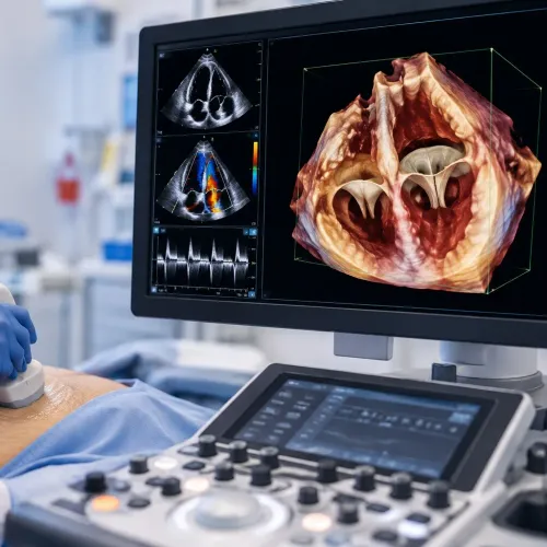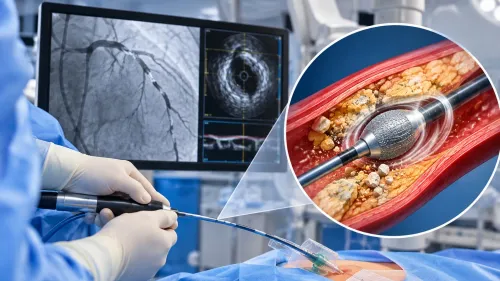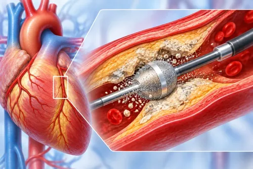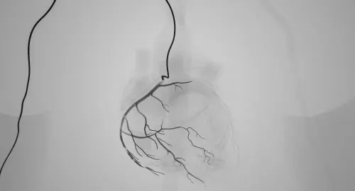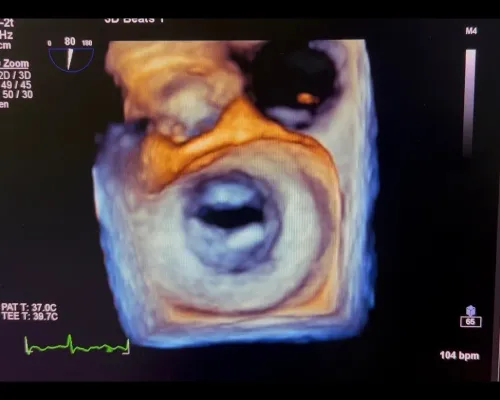
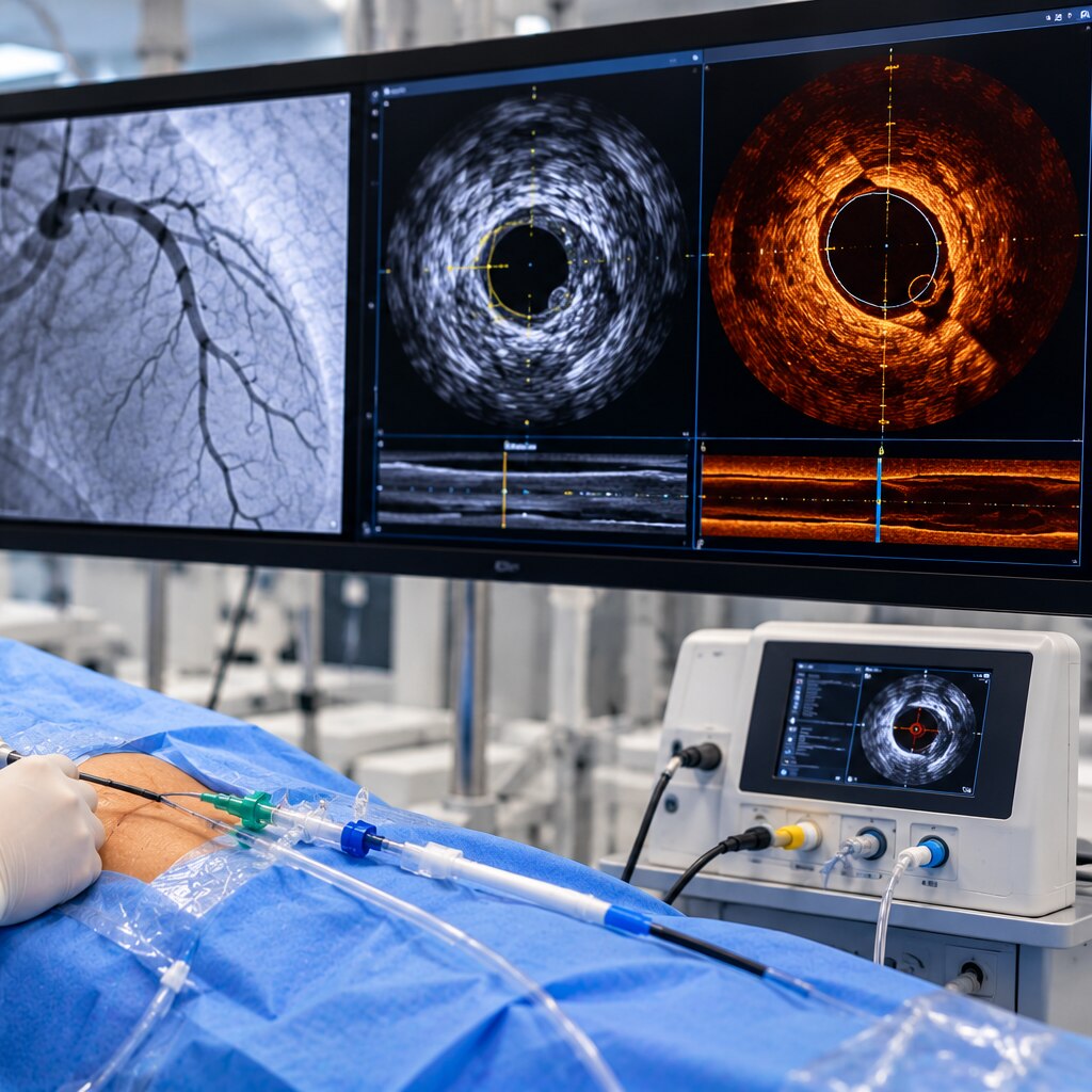
A coronary angiogram shows the artery’s contrast-filled channel as a two-dimensional silhouette, so plaque, vessel-wall disease, and stent problems can remain hidden. By the end, you will understand how IVUS and OCT create cross-sectional views, when each method is useful, and how clinicians use the findings during coronary procedures.
Key takeaways
- IVUS uses sound waves; OCT uses light to image the artery from inside.
- OCT shows finer detail, while IVUS penetrates deeper and handles calcium better.
- Intravascular imaging can clarify vessel size, plaque, calcium, and stent expansion.
- Use IVUS or OCT when angiography cannot answer a critical procedural question.
How IVUS and OCT Create Cross-Sectional Images Inside an Artery
IVUS and OCT create cross-sectional images by scanning from inside the coronary artery, not by photographing it from outside. The IVUS meaning is intravascular ultrasound: a catheter-mounted probe sends sound waves through the vessel and returns images of the lumen, vessel wall, plaque, calcium, and surrounding structure.
The OCT meaning is optical coherence tomography; near-infrared light produces sharper images of the vessel surface and stent region.
| Modality | How it forms the image | What it shows |
|---|---|---|
| IVUS | Sound waves from a catheter-mounted probe | Lumen, vessel wall, plaque, calcium, and surrounding structure |
| OCT | Near-infrared light | High-resolution vessel-surface detail and stent position |
| Angiography | Contrast dye and X-rays | A two-dimensional outline, or silhouette, of the lumen |
- A guidewire crosses the coronary artery during angiography or PCI.
- The imaging catheter advances over the wire across the area of interest.
- The system records a motorised pullback, creating sequential cross-sections along the artery.
- The catheter is withdrawn, and the operator reviews the recorded images.
The OCT meaning difference from angiography is crucial: OCT shows structures from inside the vessel rather than only a dye-filled outline. Neither technique creates a simple photograph. Guidewire shadowing, motion, calcium-related signal loss, reverberation, blooming, residual blood, and catheter distortion require trained interpretation.
IVUS Versus OCT: Depth, Resolution, Blood Clearance, and Calcium
The IVUS versus OCT choice depends on the question: IVUS gives a deeper, wider view, while OCT gives a sharper view of the surface and stent.
| Modality | Resolution and depth | Most useful view |
|---|---|---|
| IVUS | About 100–200 micrometres; 4–8 mm imaging depth | Large vessels, deep or extensive plaque, total plaque burden, vessel-size measurement, and anatomy difficult to clear of blood |
| OCT | Roughly 10–20 micrometres; 1–2 mm imaging depth | Superficial calcium, individual stent struts, tissue coverage, malapposition, edge dissection, thrombus, and other stent-surface findings |
IVUS is usually the better choice when you need the external vessel boundary, the full plaque thickness, or a reliable vessel-size measurement. Its deeper penetration helps assess large arteries and lipid-rich or extensive plaque that extends beyond OCT’s limited range. IVUS can also image through flowing blood, which helps when temporary blood clearance is difficult.
OCT resolution is substantially higher, but red blood cells scatter light. OCT therefore needs OCT blood clearance with contrast or another flushing medium during the pullback. That adds contrast exposure and can make OCT less attractive when kidney risk or the planned contrast load is high.
OCT may miss the full depth of a large or lipid-rich plaque and may not show the external vessel boundary.
For calcium, OCT better defines superficial calcium’s thickness and circumferential extent. IVUS is more useful when calcium lies deep, the vessel is large, or blood clearance is impractical. Neither modality is automatically superior; the useful view is the one that answers the procedural question.
What Intravascular Imaging Reveals When the Angiogram Is Ambiguous
An angiographic silhouette fails when an intermediate coronary stenosis has an uncertain effect, the left main coronary artery is involved, lesions overlap, plaque spreads diffusely, or positive remodeling preserves the visible lumen despite substantial disease. Angiography limitations also matter when the vessel’s true size is unclear or disease continues beyond the apparent angiographic edge.
Intravascular imaging can clarify:
- Lumen area and diameter at the narrowing
- Reference-vessel diameter for comparison and sizing
- Plaque burden measurement, including disease hidden by remodeling
- Calcium location, thickness, and distribution
- Thrombus, dissection, and plaque extending beyond the visible edge
IVUS usually shows deeper vessel-wall structure and the external vessel boundary; OCT provides finer detail near the lumen but can miss deep or lipid-rich plaque. Neither replaces clinical judgment. A severe-looking plaque or small lumen does not alone prove that the lesion causes ischemia.
Interpret the images alongside symptoms, ECG changes, troponin results, angiography, and physiology such as fractional flow reserve or instantaneous wave-free ratio. No single minimum-lumen-area cutoff applies safely to every vessel or lesion because vessel size, location, reference dimensions, and measurement method differ.
Left-main decisions require vessel size, lesion location, clinical context, and physiologic assessment rather than one number.
How IVUS and OCT Help Select and Check a Coronary Stent
The operator uses IVUS-guided PCI or OCT-guided PCI to measure the reference-vessel diameter, lesion length, plaque distribution, and calcium before choosing stent diameter and length. The stent should cover diseased tissue while extending into healthy landing zones; otherwise geographic miss leaves untreated plaque at an edge.
Calcium may require lesion preparation before the stent can expand fully.
| Imaging finding | What it shows | Possible response |
|---|---|---|
| Minimum stent area | The smallest open cross-sectional area inside the stent | Additional balloon expansion for underexpansion |
| Stent malapposition and underexpansion | Struts separated from the wall or a compressed stent | High-pressure balloon treatment or, rarely, revised treatment |
| Edge dissection or residual disease | Injury or plaque at a stent edge | Extend treatment or place another stent when necessary |
| Tissue protrusion or thrombus | Material bulging through or collecting around struts | Further assessment and treatment based on severity |
After deployment, imaging checks stent optimization rather than trusting the angiographic silhouette. IVUS measures deep vessel dimensions and plaque; OCT resolves individual struts, malapposition, tissue protrusion, thrombus, edge dissection, and disease behind struts or at the edges. These findings can prompt another balloon inflation or a different treatment before the procedure ends.
Evidence supports careful use, not guaranteed success. ULTIMATE reported 3-year target-vessel failure of 6.6% with protocol-guided IVUS implantation versus 10.7% with angiography guidance. ILUMIEN IV found greater minimum stent area with OCT, but no statistically significant reduction in its primary 2-year target-vessel-failure outcome.
When Adding IVUS or OCT Is a Sensible Procedural Decision
Choose intravascular imaging when it will answer a specific procedural uncertainty, such as the vessel’s true size, plaque extent, calcium, or whether a stent is fully expanded. IVUS and OCT are adjuncts performed during the same invasive coronary angiography or angioplasty procedure, not stand-alone screening tests.
The imaging catheter advances through the coronary artery over a guidewire, crosses the area of interest, records a pullback, and is withdrawn. These added wire and catheter steps create intravascular imaging risks, including spasm, dissection, thrombosis, ischemia, and catheter-induced distortion. Ask about IVUS procedure safety in the context of your anatomy and planned treatment.
| Choice | Main trade-off | When it may fit |
|---|---|---|
| IVUS | Images through flowing blood and adds little or no contrast | Large or deep disease, unclear vessel size, or a contrast-minimisation strategy |
| OCT | Provides sharper stent and surface detail but needs a contrast flush to clear blood | Stent expansion, malapposition, edge injury, or thrombus when kidney risk is acceptable |
OCT contrast kidney risk deserves particular attention with chronic kidney disease, diabetes, dehydration, or a high planned contrast load. Ask these questions for an interventional cardiologist:
- Which uncertainty will imaging resolve?
- Does IVUS or OCT fit my vessel and kidney-risk profile?
- How would the result change stent selection, balloon expansion, or further treatment?
A consultation with Dr. Rajesh Matta can weigh those answers alongside your angiogram, symptoms, ECG, laboratory findings, and long-term cardiac plan.
Frequently asked questions
How do IVUS and OCT create images inside a coronary artery?
A catheter-mounted IVUS probe sends ultrasound waves through the artery. OCT uses near-infrared light to create high-resolution cross-sectional images of the lumen, plaque, vessel wall, and stent.
What is the difference between IVUS and OCT?
OCT provides finer image resolution, while IVUS penetrates deeper into the vessel and visualises heavily calcified plaque more effectively. OCT also requires blood clearance for imaging.
What can IVUS or OCT reveal when an angiogram is ambiguous?
They can show the true vessel diameter, plaque burden, calcium, lesion length, dissection, and the cause of an unclear or unexpected angiographic appearance.
How do IVUS and OCT help with coronary stents?
They help select stent size and length, then check stent expansion, apposition, edge problems, and tissue or plaque that remains inside the treated segment.