Coronary angiography is most useful when the likelihood of a clinically important blockage is high enough that seeing the artery directly could change treatment. You will learn when it is urgent, when a non-invasive test is better first, how doctors interpret the results, and what to expect before and during the procedure.
Key takeaways
- ST-elevation myocardial infarction requires immediate angiography and primary PCI when appropriate.
- Use coronary CT or stress testing first when symptoms are stable and urgent blockage is unlikely.
- Doctors weigh symptoms, ECG changes, troponin results, kidney function, bleeding risk, and test findings.
- A blockage on angiography does not automatically mean you need angioplasty or a stent.
When is coronary angiography an urgent test?
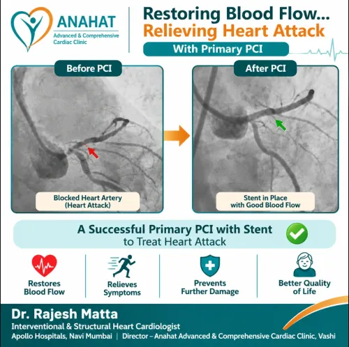
Persistent chest pain with evidence of active heart damage is not an outpatient-testing problem. Suspected ST-elevation myocardial infarction requires immediate coronary angiography so doctors can open the artery with primary PCI when appropriate; treatment should not wait for a treadmill test or coronary CT.
NSTEMI and unstable angina require hospital assessment, with urgency determined by instability, ECG changes, troponin levels, and ongoing symptoms.
- Arrange immediate angiography for shock, very low blood pressure, cardiac arrest, life-threatening ventricular arrhythmia, acute heart failure caused by ischemia, or recurrent ischemic ECG changes.
- Treat recurrent or persistent chest pain despite medication as a warning of persistent ischemia, especially when the ECG shows new ST-segment depression or T-wave changes.
- Use an early invasive strategy during the admission for high-risk NSTEMI or unstable angina, such as a definite troponin rise or fall, dynamic ECG changes, reduced heart function, diabetes, kidney disease, or previous coronary disease.
- Do not send a stable patient straight to catheterization because of a positive stress test alone; confirm that symptoms, test severity, and the likely treatment decision justify it.
| Situation | Usual timing | Why |
|---|---|---|
| ST-elevation myocardial infarction | Immediate | Reperfusion may limit heart-muscle loss |
| Very-high-risk NSTEMI or unstable angina | Immediate | Ongoing ischemia or instability threatens life |
| Other high-risk NSTEMI | During hospital admission | Early angiography guides treatment |
| Stable symptoms or an isolated abnormal stress test | Outpatient evaluation | CT or another non-invasive test may clarify risk |
Angiography carries bleeding, artery injury, contrast-associated kidney injury, stroke, and rare death risks, so doctors reserve it for cases where defining the arteries could change treatment.
When should a non-invasive test come first?
Stable, lower-risk symptoms usually justify a non-invasive test before catheter-based angiography. Coronary CT angiography is often the preferred first anatomical test for people without known coronary disease and with low-to-intermediate likelihood of obstructive disease. It can rule out significant narrowing without placing a catheter in an artery.
| Option | What it shows | Main limitation |
|---|---|---|
| Coronary CT angiography | Coronary anatomy and plaque; useful for excluding obstructive disease | Contrast, radiation, kidney impairment, heavy calcium, or irregular heart rate can reduce image quality |
| Treadmill stress testing | Symptoms, exercise capacity, ECG changes, and blood-pressure response during exertion | Less useful when the resting ECG is abnormal or you cannot exercise adequately |
| Stress echocardiography | Heart-wall movement during exercise or medication-induced stress | Image quality can limit interpretation |
| Nuclear stress testing | Blood-flow differences between stressed and resting heart muscle | Involves radiation and can produce false-positive or equivocal results |
| Invasive angiography | Direct images of the artery lumen, with a route to angioplasty if needed | Arterial access, contrast exposure, bleeding, and rare serious complications |
A positive stress test does not automatically require invasive angiography. Doctors weigh the result against your symptoms, exercise capacity, ECG, estimated pre-test likelihood, and whether finding a blockage would change treatment; an equivocal result can be clarified with another non-invasive test.
Catheter angiography becomes more reasonable with severe or persistent symptoms, high-risk imaging findings, strongly suggestive symptoms despite a negative test, or a serious possibility of revascularisation. Coronary calcium, diabetes, family history, or an abnormal resting ECG alone does not usually justify catheterisation.
How do doctors decide whether angiography is justified?
Angiography is justified when the chance of a clinically important coronary narrowing is high enough that defining the arteries could change treatment. Doctors estimate this pre-test probability from symptoms, age, examination, risk factors, ECG, troponin, and known coronary disease—not from one finding alone.
- Ongoing or recurrent chest pressure, breathlessness, or reduced exercise tolerance despite treatment raises concern.
- Dynamic ST-segment or T-wave changes on an ECG, or a raised or changing troponin, strengthens concern for acute coronary disease.
- Previous heart attack, stent, bypass surgery, or known coronary narrowing increases the likelihood that new symptoms reflect obstructive disease.
- Diabetes and kidney disease increase cardiovascular risk and influence contrast safety, but neither finding alone justifies catheterization.
- Reduced heart-pumping function or a high-risk stress-test result makes anatomical definition more useful.
A positive stress test does not automatically require angiography. Doctors consider the result’s severity, your symptoms, exercise capacity, ECG, and whether revascularization is a realistic treatment; an equivocal or conflicting result may call for another non-invasive test.
For stable symptoms without known coronary disease, coronary CT angiography often comes first when clinical likelihood is low to intermediate. Invasive angiography becomes more reasonable with very high likelihood, severe symptoms despite medicines, high-risk imaging, or anatomy that may need treatment.
It is not a screening test for asymptomatic people solely because of diabetes, calcium, family history, or an abnormal resting ECG.
What should you expect before and during coronary angiography?
Before elective angiography, follow the fasting instructions you receive and arrange transport home if sedation is used. Bring a complete medicine list, especially anticoagulants, antiplatelet drugs, diabetes medicines, and allergy details. Do not stop prescribed medication without explicit instructions.
Before you sign consent, confirm that the team has checked:
- Kidney function through recent creatinine and estimated glomerular filtration rate results.
- Any previous contrast reaction, including rash, wheezing, facial swelling, or low blood pressure.
- Bleeding risk, anemia, platelet count, and use of warfarin, apixaban, rivaroxaban, or similar medicines.
- Pregnancy possibility, when relevant.
- Whether radial access at the wrist or femoral access at the groin is safest for your anatomy and planned procedure.
You receive local anaesthetic before a thin catheter enters the artery. Radial access usually causes fewer bleeding complications; femoral access remains useful when the wrist artery is unsuitable or the procedure requires it. You may feel pressure at the entry site, but the catheter should not cause sharp pain.
Contrast can produce a brief hot flush, metallic taste, or urge to urinate.
Ask what symptoms to report immediately, including severe chest pain, breathlessness, itching, or swelling. Risks include a bruise or bleeding, arterial injury, contrast reaction, kidney injury, abnormal rhythm, stroke, heart attack, and very rarely death. The expected diagnostic benefit must outweigh those risks.
What does angiography show, and does a blockage mean angioplasty?
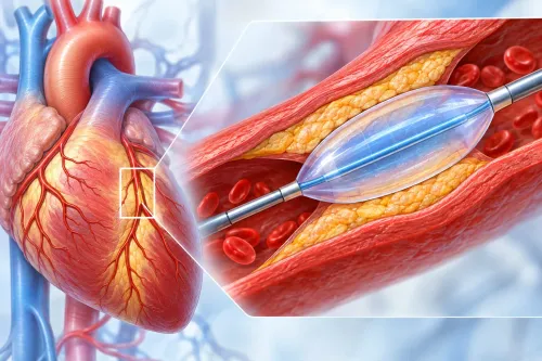
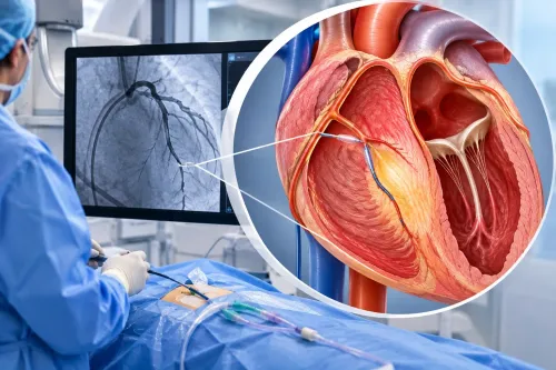
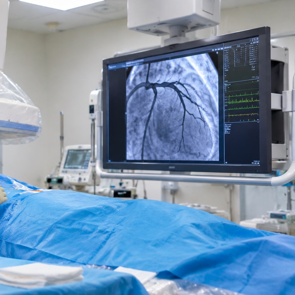
Angiography shows the location and apparent severity of narrowing inside the coronary arteries, but a tight-looking lesion is not automatically the cause of reduced blood flow. Diagnostic angiography only images the arteries; angioplasty is treatment, using a balloon and usually a stent to widen a selected blockage.
| Procedure | What it does | Decision it supports |
|---|---|---|
| Diagnostic angiography | Maps the artery lumen and narrowing | Decides whether anatomy needs further assessment or treatment |
| Angioplasty with a stent | Opens a suitable narrowing and supports the artery wall | Considered when symptoms or proven ischemia justify PCI |
| Coronary bypass surgery | Routes blood around multiple, diffuse, or surgically favourable blockages | Considered when anatomy, diabetes, heart function, or disease complexity favour CABG |
For an intermediate narrowing, doctors should not rely on visual appearance alone when there is no clear evidence of ischemia. FFR measures pressure during maximum blood-flow demand; an FFR of 0.80 or less supports a flow-limiting lesion. iFR measures pressure without giving a hyperemic drug; an iFR of 0.89 or less supports treatment.
When discussing an angiogram with Dr. Rajesh Matta, ask which lesion explains your symptoms, whether FFR or iFR is needed, and why medication, a stent, or bypass offers the best balance of benefit and risk.
In stable disease, doctors also weigh symptoms despite medical therapy, ischemia, heart-pumping function, diabetes, kidney disease, and the number and location of blockages. A blockage alone does not guarantee that revascularization will improve survival.
Related service
Coronary Angiography & Angioplasty Coronary Angiography & Angioplasty At Anahat Advanced and Comprehensive Cardiac Clinic, Dr. Rajesh Matta provides advanced care for heart blockages through Coronary Angiography and... View service → |
Frequently asked questions
When is coronary angiography an urgent test?
It is urgent when persistent chest pain accompanies evidence of active heart damage, especially suspected ST-elevation myocardial infarction. Doctors can proceed to primary PCI without waiting for a treadmill test or coronary CT.
When should a non-invasive test come first?
A non-invasive test can come first when symptoms are stable and there is no evidence of an ongoing heart attack. Doctors may choose coronary CT angiography or a stress test based on your symptoms, ECG, kidney function, heart rate, and ability to exercise.
How do doctors decide whether angiography is justified?
They combine your symptoms and examination with ECG findings, serial troponin results, previous test results, kidney function, contrast allergy history, bleeding risk, and the likelihood that finding a blockage will change treatment.
What should you expect before and during coronary angiography?
The team reviews medicines, allergies, kidney function, and bleeding risk, then places an intravenous line and prepares access through the wrist or groin. A catheter carries contrast dye to the coronary arteries while X-ray images show blood flow and narrowing.
Does a blockage on angiography mean you need angioplasty?
No. Doctors assess the blockage’s location, severity, effect on blood flow, symptoms, and overall risk. Treatment may involve medication, a stent, bypass surgery, or no procedure when the narrowing is not functionally important.