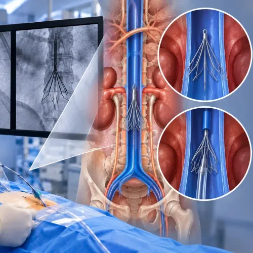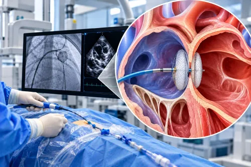
The strongest candidates are adults with non-valvular atrial fibrillation who face a meaningful embolic-stroke risk but cannot safely remain on long-term oral anticoagulation. By weighing the CHA₂DS₂-VASc score, bleeding history, heart-valve condition, anatomy, procedural risk and follow-up requirements, you can understand when closure deserves consideration and when anticoagulation remains the better-supported choice.
Key takeaways
- Candidates usually have non-valvular AF and elevated embolic stroke risk.
- Closure is considered when long-term anticoagulation is unsuitable or carries substantial bleeding risk.
- Transoesophageal echocardiography or cardiac CT checks appendage anatomy before treatment.
- You still need prescribed antithrombotic medication and follow-up imaging after closure.
Which patients are considered for left atrial appendage closure?
Adults with non-valvular atrial fibrillation, an elevated CHA2DS2-VASc score and moderate-to-high embolic stroke risk are the main left atrial appendage closure candidates.
Closure becomes worth discussing when long-term oral anticoagulation is unsafe because of major or recurrent bleeding, a non-reversible bleeding cause, or a very high bleeding risk, and you prefer a device-based strategy after counselling.
| Option | What it targets | When it fits |
|---|---|---|
| Oral anticoagulation | Clot formation throughout the circulation | Standard prevention for most eligible patients, especially when treatment is tolerated and effective |
| Left atrial appendage closure | Clot formation in the left atrial appendage | Non-valvular atrial fibrillation with suitable anatomy, stroke risk and a strong reason to avoid long-term anticoagulation |
| Warfarin-centred treatment | Clot formation throughout the circulation | Mechanical heart valves or moderate-to-severe rheumatic mitral stenosis, where closure is not an equivalent substitute |
A high CHA2DS2-VASc score identifies the need for stroke prevention; it does not prove that closure is safer or better than anticoagulation. Closure does not stop atrial fibrillation or prevent clots forming elsewhere, so another medical reason for anticoagulation can still require it.
Before proceeding, specialists assess kidney function, bleeding history, medication reliability, procedural risk, appendage anatomy and any existing thrombus with transesophageal echocardiography or cardiac CT.
How does closure compare with anticoagulant treatment?
For most eligible patients, oral anticoagulants remain the better fit because they prevent clot formation throughout the circulation without an invasive procedure.
A device-based approach is reasonable mainly for nonvalvular atrial fibrillation with meaningful stroke risk when long-term anticoagulation is contraindicated by a nonreversible bleeding cause, or when bleeding risk is high and you prefer closure after discussing the trade-offs.
| Option | What it does | Better fit | Main limitation |
|---|---|---|---|
| Direct oral anticoagulants | Reduce clotting throughout the circulation | Most eligible nonvalvular atrial-fibrillation patients | Bleeding, adherence and kidney-dose concerns |
| Warfarin | Reduces clotting throughout the circulation | Mechanical heart valves and selected valve-related conditions | INR monitoring, food and drug interactions |
| Left atrial appendage closure | Seals the appendage where many atrial-fibrillation clots form | Patients needing a device alternative because anticoagulation is unsuitable or carries high bleeding risk | Procedure complications, leaks and device-related clots |
Randomized evidence shows closure can perform comparably to direct oral anticoagulants in selected patients with high stroke and bleeding risk; it does not show that closure is safer or better for everyone. The procedure also does not stop atrial fibrillation or prevent clots from other cardiac sites.
You will usually need temporary anticoagulant or antiplatelet treatment after implantation. Follow-up imaging checks for a device-related clot or significant leak before medication is reduced. Mechanical valves and moderate-to-severe rheumatic mitral stenosis require particular caution: warfarin or another specialist-directed anticoagulation plan remains central, so closure is not an equivalent substitute.
Which medical conditions can support or rule out closure?
Closure has its strongest rationale when non-valvular atrial fibrillation creates substantial stroke risk and anticoagulation cannot be sustained. A high CHA₂DS₂-VASc score supports stroke prevention, not closure by itself.
| Finding | How it changes eligibility | What it does not mean |
|---|---|---|
| Prior spontaneous intracranial haemorrhage or severe, nonreversible gastrointestinal bleeding | Supports closure when long-term anticoagulation is unsafe | A treatable bleeding source is not a permanent reason to avoid anticoagulation |
| Kidney disease or liver disease | May increase drug exposure, bleeding risk and monitoring difficulty; assess severity and treatment options | Organ disease alone does not establish that closure is safer |
| Recurrent falls that caused serious bleeding and cannot be addressed | Supports considering closure | Fall risk without serious bleeding does not automatically rule out anticoagulation |
| Mechanical heart valve or moderate-to-severe rheumatic mitral stenosis | Usually rules out closure as an equivalent replacement for warfarin | Closing the appendage does not remove the valve-related need for anticoagulation |
A closure device only targets clot formation in the left atrial appendage. If the suspected stroke mechanism is a mechanical valve, rheumatic mitral stenosis, ventricular thrombus or arterial disease, closing the appendage will not treat that source.
Other anticoagulation needs also matter. A person who must remain anticoagulated for venous thromboembolism, a mechanical valve or another condition gains little medication-saving benefit from closure.
If anticoagulation works well without significant bleeding, closure offers less reason to accept procedural risk. The decision turns on reversible bleeding causes, organ function, anatomy, procedural risk and informed preference.
What must be checked before and after the procedure?
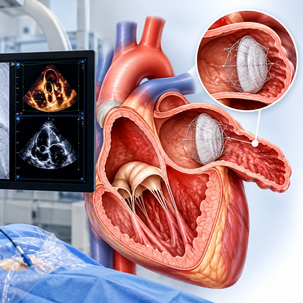
Before implantation, clinicians must confirm that the left atrial appendage contains no clot and that the device can seal it without obstructing nearby structures. A visible left atrial appendage thrombus usually requires separate specialist treatment because manipulating it can dislodge the clot.
1. Transoesophageal echocardiography or cardiac CT checks for thrombus, leaks, nearby anatomy and unusual appendage shapes. The images guide appendage anatomy and device sizing, including the opening’s width and depth. An unsuitable shape or size can make closure unsafe or incomplete.
2. The team reviews kidney function, bleeding history, access-site risks and other reasons you might still need anticoagulation. Closure does not prevent clots forming elsewhere, and it does not remove risks from atrial fibrillation or other stroke mechanisms. The procedure itself can cause pericardial effusion or tamponade, vascular bleeding, device embolization or stroke.
3. After implantation, follow-up imaging checks whether the device remains in position, whether blood flows around it, and whether device-related thrombus has formed. Transoesophageal echocardiography or cardiac CT is used according to the implant and local protocol.
Do not stop antithrombotic treatment immediately unless the treating team instructs you to do so. You will receive a temporary anticoagulant, antiplatelet treatment or both; clinicians reduce that treatment only after reviewing the imaging and your bleeding risk. A significant leak or device thrombus can require continued or altered treatment.
What should you discuss before choosing closure?
Choosing closure means trading one set of risks for another, not removing stroke risk. Discuss whether your bleeding history is reversible, whether kidney function or missed doses make anticoagulation difficult, and whether you need anticoagulation for another reason that closure cannot address.
| Plan | Main advantage | Personal cost or uncertainty |
|---|---|---|
| Continue anticoagulation | Protects against clots throughout the circulation without an invasive procedure | Ongoing bleeding risk, monitoring or dosing burden, and missed-dose consequences |
| Percutaneous closure | Targets the left atrial appendage and may reduce dependence on long-term anticoagulation | Procedure-related complications, including tamponade, vascular bleeding, embolisation or procedure-related stroke |
| Another plan | Treats a reversible bleeding source or uses a different medication strategy | It may not adequately protect you if stroke risk remains high or treatment cannot be maintained |
Ask exactly how long post-procedure antithrombotic therapy will last and what imaging must show before it is reduced. Device-related thrombus and peri-device leak can require extended treatment or another intervention.
Closure does not stop atrial fibrillation or prevent clots from elsewhere. If you already need anticoagulation for a mechanical valve, rheumatic mitral stenosis or another condition, the device may not replace it.
Dr. Rajesh Matta can help you compare your bleeding history, medication reliability, anatomy and personal tolerance for an invasive procedure rather than relying on a risk score alone.
Frequently asked questions
Which patients are considered for left atrial appendage closure?
Adults with non-valvular atrial fibrillation, an elevated CHA2DS2-VASc score and moderate-to-high embolic stroke risk are considered. The discussion is strongest when long-term oral anticoagulation is unsuitable because of serious bleeding, recurrent bleeding or another clear limitation.
How does left atrial appendage closure compare with anticoagulant treatment?
Anticoagulants reduce clot formation throughout the circulation, while closure seals the left atrial appendage, where many AF-related clots form. Closure does not remove all stroke causes and usually requires a temporary antithrombotic regimen and follow-up imaging.
Which medical conditions can support or rule out closure?
A history of major bleeding, recurrent falls with injury, bleeding-prone disease or difficulty maintaining anticoagulant treatment can support evaluation. Active infection, a clot already in the appendage, unsuitable appendage anatomy or inability to take the required post-procedure medication can rule it out.
What must be checked before and after the procedure?
Before treatment, clinicians assess stroke and bleeding risk, kidney function, current medicines, valve disease, echocardiography findings and appendage anatomy with transoesophageal echocardiography or cardiac CT. Afterward, follow-up imaging checks for device position, residual leak and device-related clot before medication changes.