A coronary angiogram can show no major blockage while your symptoms, stress test, or ECG still point to inadequate blood supply. By the end, you will understand what CoroFlow measures, how clinicians judge the reliability of its results, and when pressure, flow, and microvascular testing can clarify the cause of chest pain or breathlessness.
Key takeaways
- FFR measures epicardial pressure loss, not every form of microvascular dysfunction.
- CFR assesses flow reserve; IMR estimates resistance within the coronary microcirculation.
- Validate pressure, temperature and flow signals before interpreting CoroFlow results.
- Use coronary function testing when symptoms persist despite unobstructed or treated arteries.
Why angiography and FFR do not answer every coronary physiology question
An intermediate narrowing on coronary angiography does not reveal how much it restricts blood flow, while a normal-looking artery does not prove that the coronary microcirculation works normally. FFR, iFR and RFR answer whether pressure falls across an epicardial stenosis; they do not fully explain impaired flow reserve, raised microvascular resistance or vasospasm.
| Test | Main question | Common interpretation |
|---|---|---|
| Coronary angiography | What does the artery look like? | Shows anatomy but estimates functional severity imperfectly |
| FFR | Does hyperaemic pressure fall across a lesion? | FFR ≤0.80 supports a flow-limiting epicardial stenosis |
| iFR or RFR | Is resting pressure lower beyond a lesion? | iFR/RFR ≤0.89 supports a flow-limiting epicardial stenosis |
| CoroFlow | How do pressure and coronary flow behave? | Adds CFR and IMR measurements to the angiographic picture |
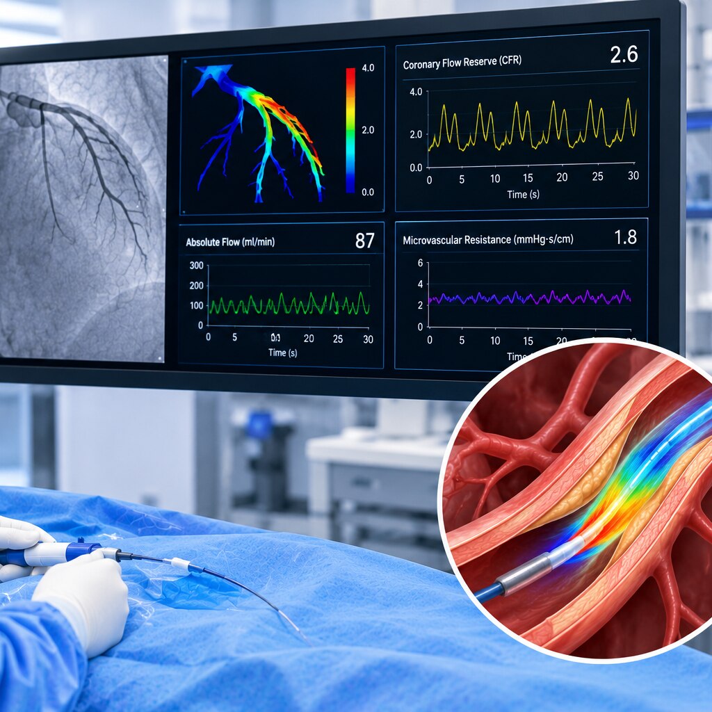
CoroFlow therefore adds information when an intermediate lesion has an uncertain pressure result, and when symptoms continue despite no obstructive coronary artery disease. Its CFR compares resting flow with flow during drug-induced hyperaemia; IMR estimates resistance in the small coronary vessels.
A normal CFR does not rule out a focal stenosis, so flow and pressure results must be read together. Conversely, normal adenosine-based measurements do not exclude vasospasm, which requires separate testing such as acetylcholine provocation.
What CFR and IMR reveal about coronary blood flow
A CFR below about 2.0 is generally considered abnormal. Coronary flow reserve compares resting coronary flow with flow during adenosine-induced maximal hyperaemia, showing how much additional blood-flow capacity remains. A low CFR can indicate impaired vasodilator capacity, but it does not identify the location of the problem.
A reduced CFR can result from:
- A flow-limiting epicardial stenosis
- Diffuse coronary atherosclerosis
- Microvascular dysfunction
- High resting flow caused by tachycardia, hypertension or increased ventricular workload
- Inadequate pharmacological hyperaemia
The index of microvascular resistance, or IMR, estimates resistance within the coronary microcirculation during hyperaemia. It combines distal coronary pressure with hyperemic transit time, so epicardial narrowing affects it less than CFR. An IMR 25 or higher is the commonly used threshold for increased microvascular resistance.
Treat these thresholds as physiology findings, not standalone diagnoses. An IMR below 25 does not exclude endothelial dysfunction or vasospasm, because adenosine primarily tests vasodilator capacity; acetylcholine testing assesses endothelial-dependent constriction and spasm. Poor adenosine delivery, recent caffeine or methylxanthine exposure, hypotension, or an inadequate hyperemic response can distort both measurements.
Interpret CFR and IMR alongside symptoms, angiography, pressure data and test quality.
How clinicians collect and validate CoroFlow measurements
Before recording, the clinician calibrates the CoroFlow system and prepares both wires under stable catheter conditions. The Doppler flow wire is advanced into the target coronary artery and positioned for a clear, aligned velocity signal; the pressure wire is zeroed to atmospheric pressure and equalized with the guide catheter.
- Confirm that the guide catheter is not obstructing the vessel or damping aortic pressure.
- Check the Doppler signal at rest, repositioning the Doppler flow wire if the waveform is weak, intermittent, or misaligned with flow.
- Record resting pressure and flow only after the signals remain stable.
- Check caffeine and methylxanthine exposure because these substances can blunt the response to adenosine hyperaemia.
Adenosine hyperaemia follows through the selected delivery route, with the clinician watching for a stable peak response rather than accepting the first numerical reading. Inadequate drug delivery, hypotension, severe airway disease, or an incomplete response can falsely elevate FFR, reduce measured CFR, and underestimate microvascular resistance.
During hyperaemia, the operator records distal coronary pressure and Doppler flow velocity together. Repeated runs strengthen confidence when values vary. The pressure wire is then checked for pressure-wire drift by returning it to the guide-catheter tip and comparing distal and aortic pressures; a mismatch requires drift correction or repeat measurement.
A credible result therefore depends on calibration, signal quality, stable hyperaemia, and documented drift verification—not the displayed number alone.
How the results distinguish epicardial and microvascular disease
An FFR of 0.80 or less with a pressure drop at one discrete lesion supports flow-limiting focal epicardial coronary disease. A gradual pressure decline on pullback points instead to diffuse atherosclerosis, especially when no single narrowing explains the pressure loss.
| Pattern | What the findings suggest | How clinicians separate it |
|---|---|---|
| Focal epicardial disease | Abnormal FFR at a discrete stenosis; CFR may also be reduced | The focal pressure gradient identifies the flow-limiting lesion |
| Diffuse disease | Gradual pressure loss, reduced CFR, and no dominant stenosis | Low CFR with preserved or only mildly raised IMR suggests impaired flow through diffuse epicardial disease, though microvascular dysfunction can coexist |
| Coronary microvascular dysfunction | CFR below the laboratory threshold or IMR 25 or higher, with no obstructive epicardial explanation | Raised IMR indicates increased microvascular resistance; low CFR alone cannot distinguish this from diffuse atherosclerosis |
| Endothelial dysfunction | Acetylcholine provokes abnormal vasoconstriction or reduced flow instead of healthy vasodilation | The response indicates endothelial-dependent dysfunction; adenosine tests vasodilator capacity and cannot exclude it |
| Epicardial or microvascular spasm | Acetylcholine reproduces angina and ischaemic ECG changes, with epicardial constriction for epicardial spasm or no visible epicardial spasm for microvascular spasm | A normal adenosine result does not rule out vasospastic disease |
The microvascular angina diagnostic criteria require compatible symptoms or ischaemia, evidence of impaired microvascular function, and no obstructive epicardial cause. Acetylcholine testing supplies complementary information rather than replacing adenosine-based CFR and IMR measurements.
When advanced coronary function testing can change the next decision
After angiography, CoroFlow is most useful when symptoms persist but the next step is uncertain: an intermediate narrowing needs functional assessment, or the angiogram shows no obstructive coronary artery disease.
In patients with angina with no obstructive coronary artery disease, combining symptoms, ECG or imaging evidence, CFR, IMR, and acetylcholine responses can identify a treatable endotype rather than leaving the cause labelled as “non-cardiac.”
A physiology-defined result can change the plan:
- A flow-limiting epicardial lesion supports revascularisation decisions; a non-flow-limiting lesion supports medical treatment instead of stenting.
- Reduced CFR or IMR of 25 or higher, with compatible symptoms and no better explanation, supports microvascular angina. Treatment may include anti-anginal therapy, blood-pressure and lipid control, and risk-factor management.
- Acetylcholine-provoked epicardial or microvascular spasm directs treatment towards vasospasm management. A normal adenosine response does not exclude it.
The CorMicA trial found that treatment guided by invasive coronary-function testing improved angina and quality of life at one year compared with usual care. Dr. Rajesh Matta can help place such results alongside ventricular function, filling pressures, anaemia, fever, blood pressure, and medication use before changing treatment.
Follow-up should track symptoms and exercise tolerance, not repeat an isolated number without a clinical reason.
Frequently asked questions
Why do angiography and FFR not answer every coronary physiology question?
Angiography shows the artery’s appearance, while FFR, iFR and RFR assess pressure changes across an epicardial narrowing. They do not fully measure coronary flow reserve, microvascular resistance or vasospasm.
What do CFR and IMR reveal about coronary blood flow?
CFR compares coronary blood flow at rest with flow during maximal vasodilation. IMR estimates resistance in the coronary microcirculation during hyperaemia, helping identify impaired microvascular function.
How are CoroFlow measurements collected and validated?
Clinicians record coronary pressure and temperature signals during catheter-based testing, obtain resting and hyperaemic measurements, and check signal quality and physiological consistency before interpreting CFR or IMR.
How can results distinguish epicardial and microvascular disease?
A pressure-based result can indicate an epicardial flow-limiting stenosis, while abnormal CFR or elevated IMR with no significant epicardial obstruction points toward microvascular dysfunction. Vasospasm requires appropriate provocative assessment.
When can advanced coronary function testing change the next decision?
Testing can clarify persistent angina after a non-obstructive angiogram, symptoms after stent treatment, or discordant angiography and pressure results. The findings can guide medical therapy, further assessment or treatment of an epicardial lesion.