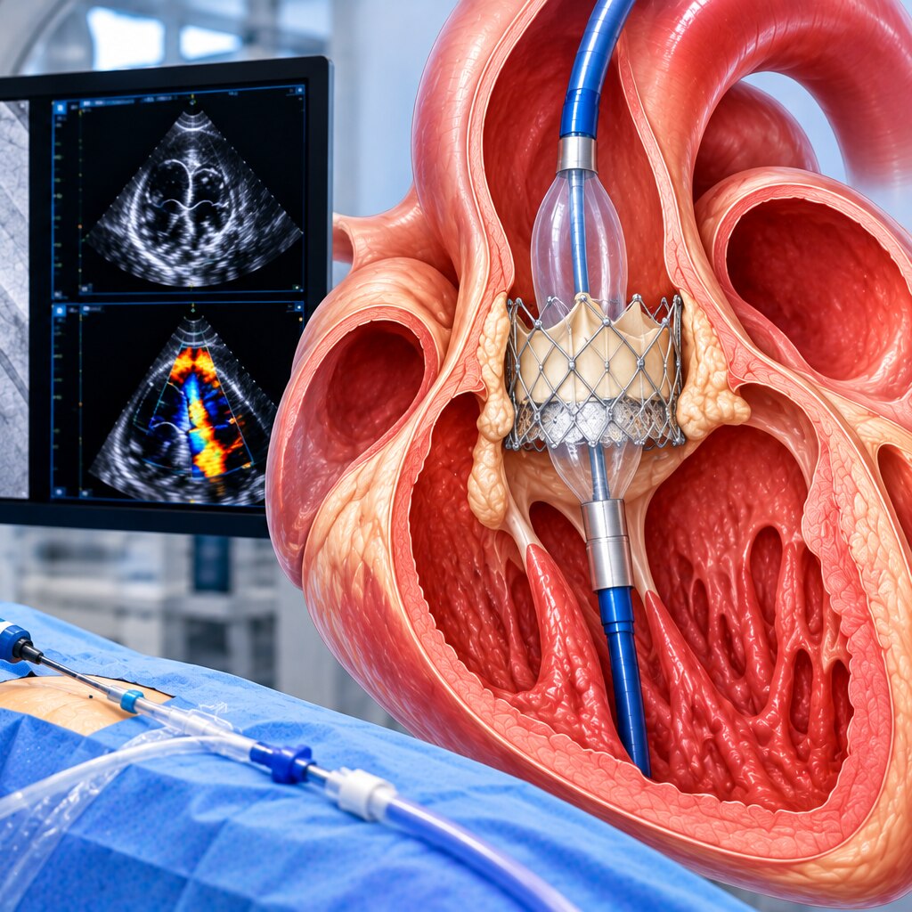
Severe aortic stenosis does not automatically mean that TAVI/TAVR is the right next step. You will learn how symptoms, echocardiography, heart function, life expectancy and valve anatomy determine whether to monitor the condition, replace the valve, or compare TAVI with surgical replacement.
Key takeaways
- Confirm severity with echocardiography, valve area, flow and pressure measurements.
- Symptoms or heart strain from severe stenosis usually prompt valve replacement evaluation.
- A Heart Team compares TAVI with surgery using anatomy, age, risk and durability.
- CT imaging and lifetime valve planning can change which replacement option fits.
What severe aortic stenosis does to the heart
Severe aortic stenosis means the aortic valve has become tight enough to obstruct blood leaving the left ventricle. This aortic valve narrowing forces the muscle to pump harder; over time, the left ventricle can thicken, weaken, or fail.
Echocardiography often defines severe disease as a valve area of 1.0 cm² or less, a peak jet velocity of at least 4.0 m/s, or a mean gradient of at least 40 mmHg, interpreted together rather than as a single decisive number.
| Pattern | What it means | What to do |
|---|---|---|
| Asymptomatic severe stenosis | Severe measurements without valve-related symptoms or damage | Arrange cardiology follow-up and repeat testing; do not assume replacement is immediately needed |
| Symptomatic severe stenosis | The valve is likely limiting blood flow during activity | Seek prompt assessment for treatment planning |
| Severe stenosis with heart damage | Reduced left-ventricular pumping function, even without clear symptoms | Seek prompt specialist assessment because waiting can allow further damage |
Report exertional breathlessness, reduced exercise tolerance, chest pain, dizziness, fainting or near-fainting promptly. Ankle swelling, breathlessness when lying flat, or breathlessness at rest suggests congestion and needs urgent medical assessment.
Symptoms can have other causes, so clinicians must establish that the valve—not lung disease, anaemia, rhythm disturbance, or coronary disease—is responsible before recommending TAVI/TAVR or another valve replacement.
How tests confirm whether stenosis is truly severe
A concordant pattern supports severe stenosis: peak jet velocity of at least 4.0 m/s, mean pressure gradient of at least 40 mmHg, and an aortic valve area of 1.0 cm² or less. Transthoracic echocardiography measures these values together with left-ventricular ejection fraction and stroke volume.
| Measure | Finding supporting severe stenosis | Why it needs context |
|---|---|---|
| Peak jet velocity | At least 4.0 m/s | High blood flow can raise the velocity |
| Mean pressure gradient | At least 40 mmHg | Low flow can produce a deceptively low gradient |
| Aortic valve area | 1.0 cm² or less | The result depends on accurate Doppler and outflow-tract measurements |
One number can mislead because velocity and gradient depend on how much blood crosses the valve. A weak ventricle, or a normal-strength ventricle that ejects a small stroke volume, can produce a low gradient even when the valve is severely narrowed.
A small aortic valve area with a mean pressure gradient below 40 mmHg is called low-flow, low-gradient stenosis when flow is reduced. It can occur with reduced ejection fraction or preserved ejection fraction with a small stroke volume.
The clinician may repeat Doppler measurements, use dobutamine stress echocardiography when ejection fraction is reduced, or obtain CT aortic-valve calcium scoring. These checks help separate genuinely severe narrowing from an underestimated valve area or a low-flow state.
When valve replacement is considered instead of monitoring
Valve replacement becomes more appropriate than surveillance when severe aortic stenosis is causing symptoms or measurable damage to the left ventricle.
For symptomatic severe aortic stenosis, exertional breathlessness, chest pain, fainting or near-fainting should prompt assessment for valve intervention, but clinicians must confirm that the valve—not lung disease, anaemia, coronary disease or an abnormal rhythm—is causing the symptoms.
The decision also changes when testing shows the heart is struggling, even before symptoms are obvious. Key findings include:
- A left ventricular ejection fraction below 50% without another explanation
- An abnormal exercise testing result, such as breathlessness, chest pain, a fall in blood pressure or an inadequate rise in blood pressure
- Very severe narrowing, often defined by a peak aortic-jet velocity of at least 5.0 m/s
- Rapid progression on serial echocardiograms
- Markedly elevated natriuretic peptide levels that support pressure-related cardiac strain
An asymptomatic severe aortic stenosis diagnosis does not automatically require TAVI/TAVR or surgical replacement. If ventricular function is preserved, exercise testing is normal and the disease is not progressing quickly, repeated echocardiography with clinical review remains appropriate.
A mean gradient below 40 mmHg does not rule out severe disease in low-flow, low-gradient stenosis. Doctors may need repeat Doppler measurements, dobutamine stress echocardiography or CT aortic-valve calcium scoring before deciding; replacing a valve that has not been confirmed as truly severe exposes you to procedural risk without treating the cause.
How the Heart Team chooses TAVI or surgical replacement
Age sets a starting point, not the verdict. The Heart Team weighs expected survival, surgical risk, frailty, existing illness, anatomy, access route, valve durability and your priorities before recommending TAVI versus surgical aortic valve replacement.
| Factor | TAVI may fit | Surgery may fit |
|---|---|---|
| Age and life expectancy | Age over 80 or life expectancy below about 10 years, when transfemoral access is feasible | Younger patients with expected survival beyond 20 years, when long-term durability and repeat procedures matter |
| Surgical risk and illness | Frailty, previous chest radiation, a porcelain aorta, organ disease or disability that makes open surgery harder | Low surgical risk, good physical reserve and few conditions that increase recovery risk |
| Personal preference | Priority for a less invasive procedure and faster early recovery, after accepting its specific complications | Willingness to accept open surgery for established durability and easier access to the heart for future treatment |
A low STS-PROM score does not settle the decision. It can understate frailty, disability, hostile chest anatomy and serious organ disease. TAVI still carries risks such as vascular bleeding, stroke, kidney injury, coronary obstruction, valve leakage and permanent pacemaker implantation.
The Heart Team also checks whether either option is likely to improve your life. If expected survival is under one year, severe illness makes benefit unlikely, or treatment cannot improve quality of life, symptom-focused care may be more appropriate.
During an assessment with Dr. Rajesh Matta, ask which risks the score misses and how today’s choice affects future valve or coronary procedures.
Related services
TAVI / TAVR Procedure Transcatheter Aortic Valve Implantation (TAVI), also known as Transcatheter Aortic Valve Replacement (TAVR), is a minimally invasive procedure used... View service → |
Structural Heart Procedures – TAVI, MitraClip & TriClip Structural Heart Procedures – TAVI, MitraClip & TriClip At Anahat Advanced & Comprehensive Cardiac Clinic, Dr. Rajesh Matta offers advanced structural heart interventions for patients with complex valve... View service → |
When anatomy, procedural risk and lifetime planning change the decision
TAVI becomes less suitable when contrast-enhanced CT shows anatomy that makes valve positioning, sealing or future coronary access difficult. A bicuspid aortic valve, heavy or uneven calcium, a low coronary-artery origin, a small annulus or unsuitable femoral access can increase complications or shift the balance toward surgical replacement.
| Finding | Why TAVI becomes riskier or incomplete | Planning consequence |
|---|---|---|
| Bicuspid aortic valve | Uneven leaflets and calcium can cause poor expansion or paravalvular leak | Compare carefully with surgical replacement |
| Low coronary height or narrow aortic root | The implanted valve can block coronary flow, causing coronary obstruction | Model the risk before choosing a valve |
| Small annulus or heavy annular calcium | Sizing becomes difficult; rupture, leak or high residual gradients can result | Assess whether surgery offers better haemodynamics |
| Small or diseased iliofemoral arteries | Femoral access can cause bleeding, dissection or vessel injury | Consider another access route or surgery |
| Prior valve intervention | A second valve may obstruct coronaries or leave limited future options | Plan later valve-in-valve treatment before implantation |
A low surgical-risk score does not capture frailty, porcelain aorta, prior chest radiation, organ disease or disability. TAVI also carries risks including stroke, kidney injury, pacemaker requirement and bleeding.
Younger patients need a lifetime plan: the first tissue valve may eventually fail, and future coronary procedures or valve-in-valve treatment may be harder after TAVI. The Heart Team should weigh durability, follow-up and the next intervention—not only the safety of today’s procedure.
Frequently asked questions
How do doctors confirm that aortic stenosis is truly severe?
They combine echocardiography findings such as aortic valve area, peak velocity and mean pressure gradient with flow assessment, left-ventricular function and, when results conflict, additional testing.
When is valve replacement considered instead of monitoring?
Replacement is considered when severe aortic stenosis causes symptoms, reduces left-ventricular function or creates other high-risk findings. Monitoring is used when severe disease has no current indication for intervention.
How does the Heart Team choose between TAVI and surgical valve replacement?
The Heart Team compares age, surgical risk, symptoms, anatomy, access vessels, valve durability, recovery priorities and the feasibility of future valve procedures.
Why can anatomy and lifetime planning change the TAVI decision?
Aortic-root anatomy, coronary artery position, access-vessel size, bicuspid valve structure and the need for future coronary access can make TAVI more or less suitable than surgery.