Severe tricuspid regurgitation can cause swelling, abdominal fullness, fatigue and breathlessness as blood backs up into the body’s veins. Understanding how TriClip reduces leakage, who may benefit, what the procedure involves and what questions to ask can help you weigh it against medicines, surgery and other catheter-based treatments.
Key takeaways
- TriClip is considered when severe TR causes symptoms and surgery carries substantial risk.
- Echocardiography checks valve anatomy, leaflet movement and whether a clip can grasp the valve.
- Ask which symptoms, test findings and alternatives support the recommendation.
- Plan for follow-up echocardiograms, medication review and monitoring after the procedure.
How severe tricuspid regurgitation affects the right side of the heart
The tricuspid valve sits between the right atrium and right ventricle. Tricuspid regurgitation occurs when the valve does not close tightly, allowing blood to flow backward into the right atrium each time the right ventricle contracts.
Severe tricuspid regurgitation raises pressure in the veins returning blood to the heart and makes the right ventricle work harder.
Over time, the right ventricle can enlarge and weaken. Blood then backs up into the body, causing leg swelling, abdominal fullness, liver congestion, reduced appetite and breathlessness. This progression can lead to right-sided heart failure.
Similar symptoms can also come from lung disease or left-sided heart problems, so the valve leak must be assessed alongside heart imaging and the person’s functional limitations.
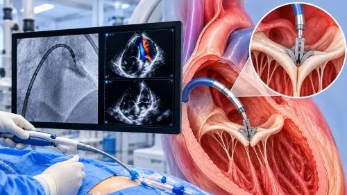
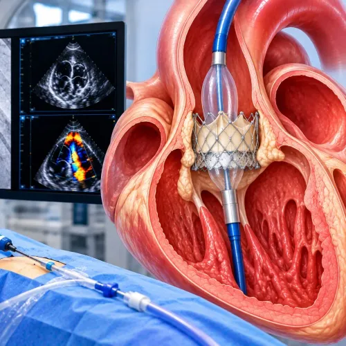
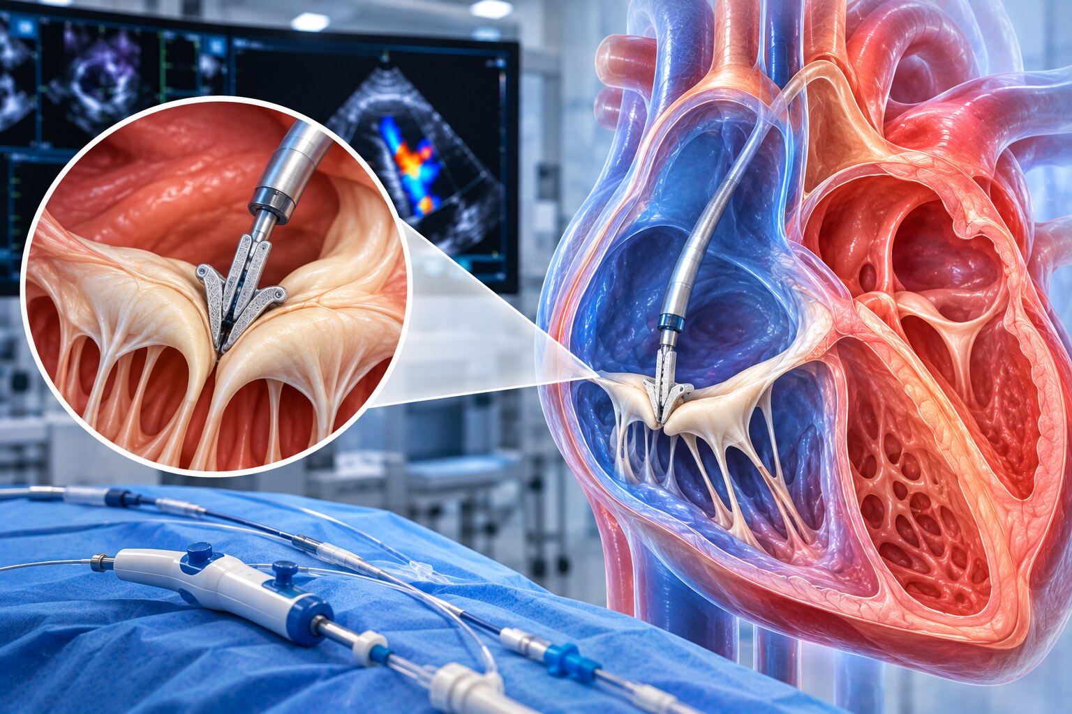
TriClip is a repair, not a valve replacement. A doctor guides a catheter through a vein, usually from the groin, into the right side of the heart. The device grasps and brings together portions of the tricuspid valve leaflets, improving their coaptation and creating smaller openings through which blood moves forward.
This reduces the backward leak, but it does not guarantee that regurgitation disappears. Reducing the leak may not restore a severely damaged right ventricle, especially when right-sided heart failure has become advanced.
How doctors decide whether TriClip is suitable
Suitability depends on more than the echocardiogram’s label. The valve team matches symptoms, leaflet anatomy, right-ventricular function, pulmonary pressure, surgical risk, and the likely benefit of reducing the leak.
| Finding | Supports TriClip | Raises concern |
|---|---|---|
| Symptoms | Breathlessness, swelling, fatigue, or abdominal fullness linked to right-sided congestion | Symptoms explained mainly by lung disease, anaemia, kidney disease, or deconditioning |
| Valve anatomy | Leaflets that the clip can grasp securely, with a targetable leak | Congenital abnormalities, severe leaflet damage, or anatomy unlike studied patients |
| Right heart | Preserved or moderately impaired right-ventricular function | Advanced irreversible right-heart failure or severe right-ventricular dilation |
| Pressure and organs | Pulmonary pressure and kidney and liver status that allow potential recovery | Severe pulmonary hypertension, marked kidney impairment, or advanced liver congestion |
| Procedural risk | Symptomatic severe TR with intermediate or greater surgical risk, particularly when surgery is unsuitable | Active endocarditis or a reasonable, safer surgical option |
The team also predicts the result after clipping. It considers how much residual TR might remain, where the remaining jet would sit, and whether the tricuspid inflow gradient would become too high. A technically successful clip is not enough if congestion and exercise limitation continue.
The evidence comes from selected patients, not every person with severe TR, massive tricuspid regurgitation, or torrential tricuspid regurgitation. A reduction in leakage cannot restore damaged right-ventricular function, so advanced heart, lung, kidney, or liver disease can limit the benefit even when the device is placed successfully.
The tests and heart-team review behind the recommendation
The recommendation rests on more than an echocardiographic severity label. Transthoracic echocardiography measures regurgitation, right-ventricular size and function, valve anatomy, pulmonary pressure and other valve disease. Transesophageal echocardiography then maps leaflet motion, the coaptation gap, jet location and pacemaker-lead interference for procedural planning.
Right-heart catheterisation clarifies pulmonary pressures and cardiac output when imaging and symptoms disagree or pulmonary hypertension is suspected. Blood tests assess kidney and liver function, anaemia and congestion.
A structural interventional cardiologist, cardiac-imaging specialist, heart-failure clinician and cardiac surgeon review the findings together. They connect breathlessness, fatigue, swelling or abdominal fullness with examination, laboratory results, lung and left-heart disease, atrial fibrillation, kidney function and deconditioning.
The team also checks for advanced right-ventricular failure, severe pulmonary hypertension, active endocarditis, congenital anatomy or leaflet features outside the studied TriClip population.
| Option | What it offers | When it applies |
|---|---|---|
| TriClip | Catheter-based reduction in leakage, without open-heart surgery | Suitable anatomy, high surgical risk, and symptoms linked to severe TR |
| Surgery | Repair or replacement, with an opportunity to address other valves | Operable patients, especially when another valve operation is needed |
| Medical treatment | Diuretics relieve congestion; treatment targets rhythm, lung, kidney and left-heart disease | Essential before and after intervention; inadequate alone when severe TR remains symptomatic |
The team must also estimate residual TR, tricuspid inflow gradient, right-ventricular function and symptoms after clipping. Trial results cannot be transferred automatically to someone with irreversible right-heart failure or markedly different anatomy.
What to expect before, during and after the procedure
Preparation usually includes a medication review, blood tests, kidney-function assessment and final echocardiographic planning. Follow the centre’s fasting instructions, arrange transport and support at home, and do not stop anticoagulants or antiplatelets unless your treating team gives a specific schedule.
1. During the procedure, doctors guide a catheter through the groin vein into the right atrium and tricuspid valve. Echocardiography directs the clip as it grasps and brings valve leaflets together. This transcatheter edge-to-edge repair creates smaller openings and reduces backward flow; it repairs the valve rather than replacing it.
Sedation or anaesthesia is used, and the procedure avoids open-chest surgery.
2. Recovery usually involves observation for bleeding, rhythm changes and kidney function, followed by gradual walking and discharge instructions. Report increasing groin swelling, fever, chest pain, fainting or worsening breathlessness promptly. Possible complications include vascular bleeding, infection, arrhythmia, stroke, kidney injury, damage to nearby structures, new tricuspid narrowing or another procedure.
3. The intended benefit is less breathlessness, swelling and fatigue, with better daily function and quality of life. TriClip does not guarantee complete elimination of the leak, and current randomized evidence has not proven longer survival or fewer heart-failure hospitalizations. Continue prescribed diuretics and treatment for conditions such as atrial fibrillation or pulmonary hypertension.
Follow-up echocardiograms and clinical reviews remain necessary.
How to assess a TriClip recommendation for your situation
A TriClip recommendation is worth accepting only if the expected gain is clear for your symptoms and daily function. FDA approval applies to symptomatic severe tricuspid regurgitation in people at intermediate or greater surgical risk with suitable anatomy; it does not confirm that clipping is the best option for you.
Ask your specialist:
1. What is causing my leak, and what do my transesophageal echocardiogram images show about leaflet motion, jet location, tethering, coaptation gap, pacemaker or defibrillator leads, and tricuspid-annular measurements?
2. How much residual tricuspid regurgitation do you expect, and could the clip create a troublesome tricuspid inflow gradient?
3. How are my right-ventricular function, kidney function, pulmonary pressures, liver congestion and swelling affecting the likely benefit and procedural risk?
4. What improvement are you targeting—less breathlessness, fewer diuretics, better walking—or merely a lower echocardiographic grade? What are the alternatives, including continued medical treatment, surgery or no procedure?
Judge the proposal against a measurable baseline. The pivotal trial showed better symptoms and quality of life at one year, not a proven survival benefit. Ask when follow-up echocardiography will check residual TR, gradients, right-ventricular function and pressures.
Dr. Rajesh Matta can use this same question-based review to help you weigh anatomy, risk and realistic goals rather than treating “successful” placement as the final result.
Related service
Structural Heart Procedures – TAVI, MitraClip & TriClip Structural Heart Procedures – TAVI, MitraClip & TriClip At Anahat Advanced & Comprehensive Cardiac Clinic, Dr. Rajesh Matta offers advanced structural heart interventions for patients with complex valve... View service → |
Frequently asked questions
How does severe tricuspid regurgitation affect the right side of the heart?
A leaking tricuspid valve sends blood back into the right atrium, increasing pressure and volume in the right-sided chambers. Over time, this can contribute to breathlessness, leg swelling, abdominal fluid retention and reduced exercise tolerance.
How do doctors decide whether TriClip is suitable?
The heart team considers symptom severity, tricuspid valve anatomy, right-ventricular function, pulmonary pressures, other heart conditions, previous treatments and surgical risk. TriClip is suitable only when the anatomy allows the device to grasp the valve leaflets safely.
What tests are used before a TriClip recommendation?
Evaluation commonly includes transthoracic echocardiography, transesophageal echocardiography, electrocardiography, blood tests and imaging that assesses the right ventricle and pulmonary circulation. The team uses these findings to judge valve anatomy, disease severity and procedural risk.
What happens before, during and after TriClip treatment?
Before treatment, doctors review medications, imaging and anaesthesia needs. During the procedure, a catheter carries the clip to the tricuspid valve through a vein, usually in the groin. Afterward, you receive monitoring, medication instructions and follow-up imaging.
How should I assess a TriClip recommendation?
Ask your cardiology team to explain the evidence of severe TR, your valve anatomy, expected benefits, important risks, alternatives and the consequences of deferring treatment. Request a clear follow-up plan with symptom checks and echocardiograms.