An IVC filter is not a routine add-on for every deep-vein thrombosis or pulmonary embolism. You will learn when anticoagulation is unsafe or ineffective enough to justify a filter, what evaluation implantation requires, and how a documented follow-up plan determines whether the device is removed or left in place.
Key takeaways
- Use an IVC filter mainly when acute DVT or PE exists and anticoagulation is unsafe.
- Review imaging, bleeding risk, kidney function, and the planned retrieval date before implantation.
- Arrange follow-up before discharge so the filter is not left in indefinitely.
- Removal uses a catheter and snare; embedded or tilted filters can require advanced techniques.
When does an IVC filter meet the clinical threshold?
The threshold is an objectively confirmed acute deep-vein thrombosis or pulmonary embolism when anticoagulation cannot safely be given. That is the main answer to when is an IVC filter recommended: the patient has an IVC filter contraindication to anticoagulation, such as active major bleeding, recent high-risk surgery, or an exceptionally high bleeding risk.
Clinicians may also consider a filter after serious bleeding caused by anticoagulants or objectively confirmed recurrent venous thromboembolism despite therapeutic treatment. Before calling treatment failure, they check:
- Adherence and the prescribed dose
- Drug interactions and absorption problems
- Whether kidney or liver changes affected drug levels
- Whether repeat imaging truly confirms new clot
- Whether the recurrent pulmonary embolism despite anticoagulation is a correct diagnosis
A routine, low-risk DVT or PE that can be treated safely with anticoagulation usually does not justify adding a filter. Prophylactic placement in someone who has not developed VTE is also not routine; it is reserved for narrowly selected situations with exceptionally high clot risk and no workable preventive medication.
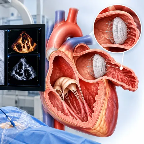
An IVC filter catches some clot travelling from the legs or pelvis through the inferior vena cava. It does not dissolve existing clot, prevent new clot formation, replace anticoagulation, or eliminate all PE risk. Start or resume anticoagulation when bleeding risk becomes acceptable.
What must be checked before implantation?
Before placement, the IVC filter pre-procedure evaluation must prove acute DVT or PE through appropriate imaging and document why anticoagulation cannot be used. Record the anticoagulant name and dose, the timing and severity of bleeding, platelet count, kidney function and contrast allergies; recent surgery, coagulopathy, infection and other conditions affecting procedural bleeding also matter.
Inferior vena cava imaging maps the clot’s location and extent, confirms IVC size and patency, and helps select the safest neck or groin venous access route. A blocked, unusually narrow or duplicated IVC can change the device or procedure.
Before consent, confirm:
- The clot imaging report and bleeding contraindication
- Kidney function and contrast imaging risk
- The selected filter, retrieval owner, review date and device details
- A contingency plan if anticoagulation remains unsafe or retrieval is not possible
| retrievable versus permanent IVC filter | Best fit | Key requirement |
|---|---|---|
| Retrievable | Temporary contraindication when later removal is plausible | Named follow-up owner and retrieval review date |
| Permanent | Long-term protection expected or retrieval risk is unacceptable | Ongoing surveillance and documented reason |
Keep the procedure report, implant card, filter model and follow-up instructions. Give them to every future clinician so nobody loses track of the device, its indication or the removal plan.
What happens during implantation and immediately afterward?
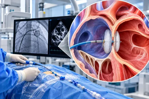
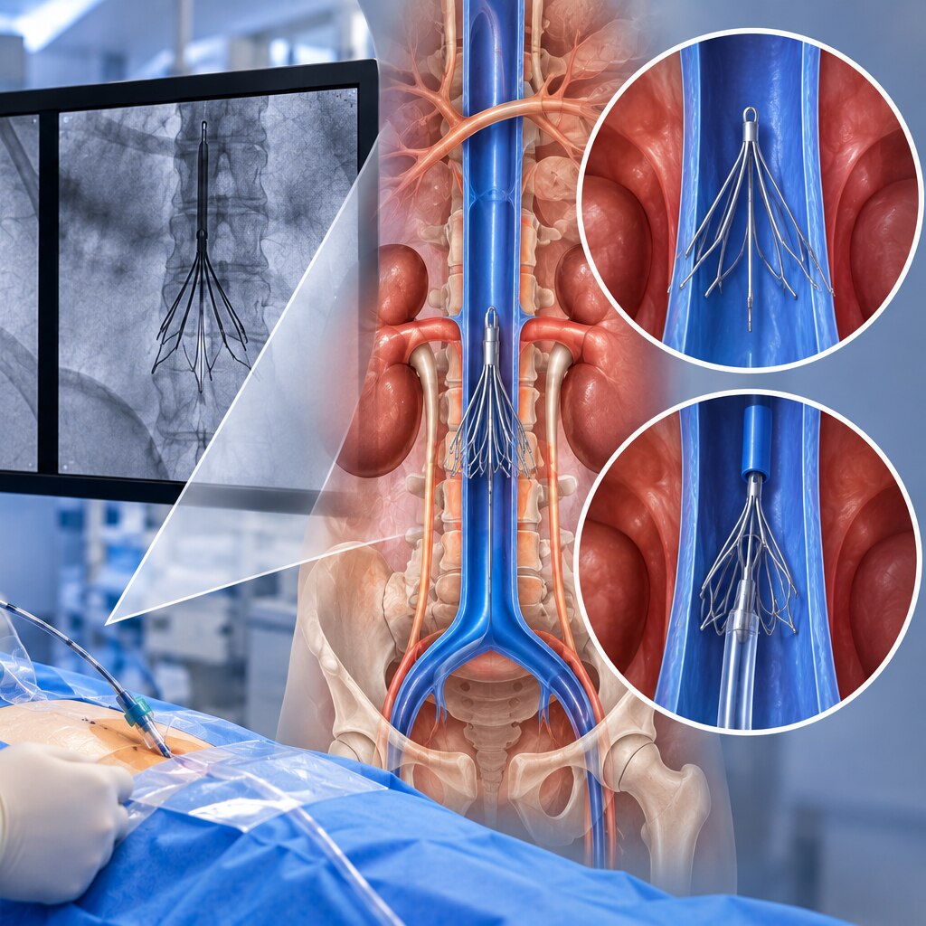
During an IVC filter implantation procedure, you receive local anaesthesia at a neck or groin venous access site. A catheter is advanced through the vein into the inferior vena cava (IVC), with fluoroscopy and contrast venography, or other imaging, used to confirm the IVC’s anatomy and the filter’s final position.
- The clinician releases the filter’s metal framework inside the IVC, where it expands against the vessel wall and remains until retrieval or indefinitely, depending on the device and plan.
- The framework intercepts some clot fragments travelling from the lower limbs or pelvis toward the lungs. It does not dissolve a clot already lodged in a leg vein or lung, prevent every pulmonary embolism, or stop new clots forming.
- You remain under observation while staff check your pulse, symptoms, and puncture site for bleeding or swelling. Follow your IVC filter recovery instructions: keep the access area clean and dry, and avoid strenuous activity or heavy lifting for the period specified by your treating team.
- Seek urgent care for uncontrolled bleeding, worsening leg swelling, chest pain, breathlessness, or fainting.
The filter does not replace anticoagulation. Your clinician continues reviewing whether and when blood-thinning treatment can start or resume, alongside any plan for clot surveillance.
How should retrieval timing and follow-up be managed?
IVC filter retrieval timing is decided by whether pulmonary-embolism protection remains necessary, not by an automatic deadline. At the follow-up consultation, the clinician reassesses when anticoagulation becomes safe, current symptoms, filter position and integrity, and any clot around or inside the device.
A named clinician or service must own the retrieval follow-up plan. The record should show the filter model, implantation date, responsible service, documented review date, and the action required after review.
- Review the filter when the original temporary contraindication to anticoagulation has resolved or the risk of pulmonary embolism has changed.
- Check whether anticoagulation can start or resume safely; the filter does not dissolve clot, prevent new clot formation, or replace anticoagulation.
- Escalate a missed IVC filter follow-up by contacting the patient, rescheduling promptly, and informing the responsible service rather than allowing the device to disappear from clinical records.
The FDA decision analysis found that removal often had a favourable benefit-risk balance around 29 to 54 days after implantation when transient PE risk had resolved. This is context, not a universal schedule; removal is considered when the temporary indication ends.
Retrieval is a separate procedure requiring imaging assessment and informed consent. If anatomy, an embedded component, trapped clot, or another hazard makes removal riskier than continued protection, the filter may remain with ongoing surveillance.
In Navi Mumbai, Dr. Rajesh Matta can review anticoagulation status and coordinate the next specialist assessment when cardiac or clot-related follow-up needs clarification.
How is removal performed, and what can make it difficult?
The IVC filter removal procedure uses catheter access rather than open surgery, but removal is not guaranteed or automatically safer than observation. Before proceeding, the clinician confirms that pulmonary-embolism protection is no longer needed, anticoagulation is feasible, and retrieval risk is acceptable.
1. Imaging checks the filter’s position and condition. Fluoroscopy, contrast venography, or cross-sectional imaging can show filter tilt and wall penetration, an embedded tip, fracture, migration, or clot trapped inside or around the device.
2. After local anaesthesia, the clinician enters a vein, usually in the neck or groin, and advances a catheter into the inferior vena cava. A retrieval sheath and snare are guided to the filter hook; the snare captures the hook, and the filter collapses into the sheath for removal.
3. Imaging before withdrawal confirms the planned route and checks for trapped thrombus. Imaging after removal confirms that the filter is out and assesses the vena cava for bleeding, injury, or residual obstruction.
4. A large thrombus, significant wall penetration, an embedded tip, fractured component, or prolonged implantation can make standard retrieval difficult or impossible. Advanced retrieval planning, another imaging review, or leaving the filter in place may be safer.
Prolonged dwell time carries recognized concerns, including IVC thrombosis or occlusion, recurrent DVT, IVC filter fracture or migration, and penetration into the IVC wall or nearby structures. Remove the filter when protection is no longer needed and retrieval risk is acceptable—not solely because a calendar date has arrived.
Frequently asked questions
When does an IVC filter meet the clinical threshold?
The main indication is objectively confirmed acute deep-vein thrombosis or pulmonary embolism when anticoagulation cannot safely be given because of active major bleeding, recent high-risk surgery, or exceptionally high bleeding risk.
What must be checked before IVC filter implantation?
The clinical team checks clot location, inferior vena cava anatomy, bleeding risk, kidney function, infection risk, imaging findings, and whether anticoagulation can begin later. They also establish a retrieval and follow-up plan.
What happens during IVC filter implantation?
A clinician guides a collapsed filter through a catheter, usually from a neck or groin vein, and releases it inside the inferior vena cava. The patient is monitored afterward for bleeding, pain, filter position, and other complications.
How should IVC filter retrieval timing and follow-up be managed?
Schedule follow-up before discharge and reassess the filter once protection from pulmonary embolism is no longer needed or anticoagulation is safe. Retrieval timing depends on the patient’s clot risk, bleeding risk, and filter type.
How is IVC filter removal performed, and what can make it difficult?
A catheter and retrieval snare remove a retrievable filter through a vein. Removal becomes more difficult when the filter tilts, its hook embeds in the vein wall, clot forms around it, or the filter fractures or penetrates the vein.