A MitraClip procedure reduces mitral valve leakage through a catheter rather than a chest incision, but it still involves advanced imaging, anaesthesia and careful hospital monitoring. By the end, you will understand how the clip works, how doctors decide whether it fits your valve and heart condition, what happens during treatment, and what follow-up involves.
Key takeaways
- MitraClip reaches the heart through a femoral vein catheter.
- TEER remains a serious procedure requiring imaging, anaesthesia, and monitoring.
- Suitability depends on valve anatomy, symptoms, heart function, and surgical risk.
- Follow-up echocardiograms show whether leakage and heart strain have improved.
How the MitraClip reduces leakage through a vein
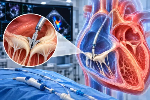
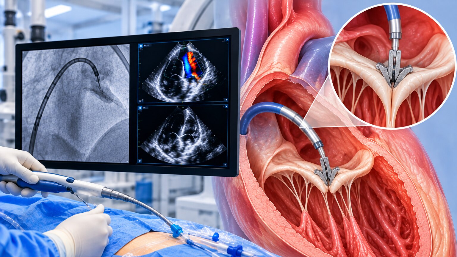
Doctors can repair the mitral valve without opening the chest by delivering a clip through a femoral vein catheter. This approach is called transcatheter edge-to-edge repair, or TEER, because it brings the two valve leaflets together at their edges rather than replacing the valve.
- The catheter enters through a vein in the groin and travels into the right side of the heart.
- Doctors guide it through the wall between the atria into the left atrium.
- Under echocardiographic and X-ray guidance, they position the MitraClip above the leaking valve.
- The clip grasps a portion of the front leaflet and a portion of the back leaflet.
- The joined leaflets form two smaller openings, limiting the gap through which blood flows backward.
This MitraClip mechanism reduces mitral regurgitation without removing the valve or using a heart-lung machine. The aim is a substantial fall in leakage while preserving enough opening for blood to pass forward. Adding clips can reduce leakage further, but too many can narrow the valve and obstruct flow.
Suitability depends on whether the leaflets have enough healthy tissue for secure grasping and whether the anatomy allows this balance.
Why avoiding open-heart surgery does not mean a minor procedure
A catheter-based repair still takes place in a fully equipped cardiac procedure room, not a routine clinic. You may receive general anaesthesia, or deep sedation in selected cases, while an anaesthesia team monitors breathing, blood pressure, heart rhythm and oxygen levels.
1. Before starting, the team reviews echocardiograms, ECG results and blood tests, places an intravenous line, and prepares the groin for venous access. A sterile sheath is inserted into the femoral vein.
2. The operator advances the delivery catheter toward the heart under fluoroscopy, which provides live X-ray guidance. Transoesophageal echocardiography supplies detailed images of the valve and guides each movement.
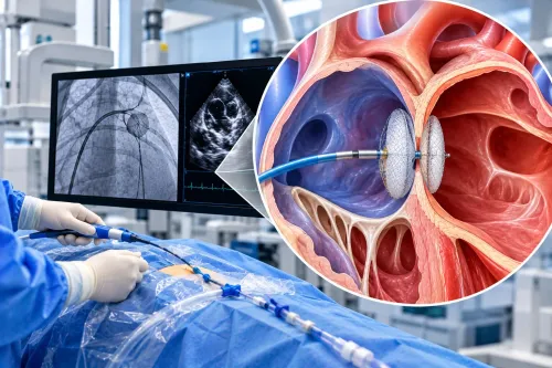
3. The catheter crosses from the right atrium into the left atrium through a controlled transseptal puncture. This step demands precise positioning because a misplaced puncture can cause bleeding, pericardial effusion or tamponade.
4. The clip is steered to the leaking valve, grasps the leaflets, and is checked with echocardiography before release. The team confirms that regurgitation has fallen without creating excessive mitral narrowing.
5. After removing the catheter, staff monitor the access site, heart rhythm, blood pressure and neurological status. Bleeding, stroke, clip detachment and residual leakage remain possible despite the absence of a chest incision.
How doctors decide whether MitraClip is suitable
Doctors recommend MitraClip when severe mitral regurgitation is causing symptoms or heart strain, and testing shows that the leaflets can be grasped without creating clinically important mitral stenosis. The assessment includes left-ventricular function and size, pulmonary pressure, surgical risk, and the valve’s exact shape.
3D transoesophageal echocardiography shows leaflet length, calcification, the valve opening, a flail leaflet or tethered leaflets, and the coaptation gap—the distance between leaflets that should meet. A large gap, heavy calcium, or inadequate leaflet tissue can prevent secure clip attachment.
| Cause | Decision focus | What must be true |
|---|---|---|
| Primary or degenerative MR | Compare durable surgical repair with TEER | Surgical risk is high or prohibitive, and the anatomy permits stable leaflet grasping |
| Secondary or functional MR | Treat heart failure and ventricular disease first | Symptoms persist despite guideline-directed therapy, with suitable anatomy and an acceptable procedure risk |
The goal is a major reduction in leakage, not always a perfectly normal valve. Additional clips can reduce residual MR but also narrow the valve and raise the transmitral gradient. Dr. Rajesh Matta can help interpret whether the scan supports TEER or whether surgery, medical treatment, or another option offers safer long-term control.
Related services
Mitraclip procedures MitraClip is a minimally invasive procedure used to treat significant mitral regurgitation (MR), a condition in which the mitral valve allows blood... View service → |
Structural Heart Procedures – TAVI, MitraClip & TriClip Structural Heart Procedures – TAVI, MitraClip & TriClip At Anahat Advanced & Comprehensive Cardiac Clinic, Dr. Rajesh Matta offers advanced structural heart interventions for patients with complex valve... View service → |
Why primary and secondary mitral regurgitation follow different pathways
The cause determines whether doctors must repair the valve itself or first treat the heart that is distorting it. In primary MR, also called degenerative MR, disease in the leaflets, chordae or supporting structures creates the leak.
In secondary MR, also called functional MR, a weakened or enlarged left ventricle pulls the valve apart; the leaflets may be relatively normal.
| Cause | Main treatment pathway | When TEER is considered |
|---|---|---|
| Primary or degenerative MR | Durable surgical repair is preferred when surgeons can achieve it at acceptable risk. | When surgical risk is high or prohibitive and the valve anatomy is suitable. |
| Secondary or functional MR | Start guideline-directed heart-failure treatment and address ventricular disease. | When severe symptoms continue despite optimized, maximally tolerated treatment. |
COAPT should not be applied to every patient with functional MR. In carefully selected COAPT patients, TEER plus medical therapy reduced death or heart-failure hospitalisation at 24 months compared with medical therapy alone: 35.8% versus 67.9%. MITRA-FR found no significant reduction, partly reflecting different patient selection, MR severity and ventricular enlargement.
Three-dimensional transoesophageal echocardiography then checks leaflet length, coaptation, flail segments, calcification and the valve opening. The clip must reduce leakage without raising the transmitral gradient enough to cause mitral stenosis.
What happens after the clip is placed
The final check weighs residual regurgitation against the transmitral gradient. Transesophageal echocardiography confirms that the clip remains attached, both leaflets move, blood flows adequately, and the valve has not become too narrow. A second clip can reduce leakage but also cause mitral stenosis.
- The team checks for bleeding or vascular injury at the femoral access site and removes the catheter once it is safe.
- Nurses monitor your blood pressure, heart rhythm, oxygen level and groin area during recovery.
- An echocardiogram before discharge assesses the clip, remaining leakage, valve gradient and heart function.
The absence of a chest incision does not eliminate serious risks. Complications include a problem from the transseptal puncture, pericardial tamponade, stroke, infection, clip detachment from one leaflet and recurrent or persistent mitral regurgitation.
Recovery is usually shorter than after surgical repair because there is no sternotomy. Expect observation overnight or a short hospital stay, with activity and medication instructions tailored to bleeding risk, heart-failure status, heart rhythm and any separate need for anticoagulation. Breathlessness may not disappear immediately if another heart or lung condition contributes to it.
How follow-up shows whether the repair is helping
Many patients leave after an overnight observation period, but recovery is judged by more than discharge timing. Early reviews check the groin puncture, bleeding, blood pressure, heart rhythm and breathlessness before activity is increased.
Follow-up echocardiography shows whether the clip still reduces leakage and whether it has narrowed the valve. Doctors compare:
- Mitral regurgitation severity and the pressure gradient across the valve
- Left-ventricular size and pumping function
- Pulmonary artery pressure
- New or recurrent symptoms, including fatigue, swelling and breathlessness
Improvement can take time. Breathlessness that persists after a technically successful repair may reflect weak left-ventricular function, pulmonary hypertension, lung disease, anaemia or atrial fibrillation rather than ongoing valve leakage.
Medication decisions depend on the reason for treatment and your other conditions. People with secondary mitral regurgitation usually continue heart-failure medicines because the clip does not replace treatment for the weakened heart. You may also need an anticoagulant for atrial fibrillation or another indication; the clip alone does not automatically require lifelong anticoagulation.
Serial examinations and echocardiograms reveal whether the benefit is lasting. Worsening symptoms or recurrent mitral regurgitation can prompt repeat imaging, medication adjustment or discussion of another intervention.
Frequently asked questions
How does MitraClip reduce mitral valve regurgitation?
A catheter carries the clip through a femoral vein to the heart, where the clip brings the mitral valve leaflets together at their edges and reduces backward blood flow.
Why is MitraClip not considered a minor procedure?
Although it avoids opening the chest, TEER requires catheter access, continuous imaging, anaesthesia or sedation, careful clip positioning, and monitoring for complications.
How do doctors decide whether MitraClip is suitable?
The heart team reviews symptoms, echocardiogram findings, valve anatomy, heart function, other illnesses, and the risks of surgery or untreated regurgitation.
Why do primary and secondary mitral regurgitation need different treatment pathways?
Primary regurgitation comes from a problem with the valve itself, while secondary regurgitation results from changes in the heart; doctors therefore address the underlying cause and assess TEER differently.
What happens after a MitraClip is placed?
Doctors check valve leakage and pressure with imaging, monitor the catheter entry site and heart rhythm, then plan medicines, discharge, activity guidance, and follow-up.