An atrial septal defect changes blood flow between the heart’s two upper chambers, but not every defect has the anatomy or pressure conditions needed for a catheter-based implant. You will learn how doctors assess suitability, guide the occluder into place, prepare you for the procedure and confirm that the heart is recovering afterward.
Key takeaways
- Secundum ASDs with suitable rims are the main defects treated by device closure.
- Echocardiography measures the defect, heart enlargement, shunt, and pulmonary pressure.
- Doctors guide the occluder through a catheter from a vein into the atrial septum.
- Follow-up imaging confirms the device position and checks for residual blood flow.
Which atrial septal defects can a closure device treat?
An atrial septal defect anatomy involves an opening in the wall separating the right and left atria. Because left-atrial pressure is usually higher, oxygen-rich blood can cross into the right atrium, creating a left-to-right shunt. Extra blood then enters the right ventricle and lungs, potentially enlarging the right heart and increasing pulmonary blood flow.
| Defect type | Location and associated anatomy | Usual treatment approach |
|---|---|---|
| Secundum ASD | Central atrial septum; the commonest type for catheter closure | Two-disc device if the defect is hemodynamically significant and has adequate rims |
| Primum ASD | Lower septum beside the atrioventricular valves; may include a cleft mitral valve | Surgical repair, including associated valve repair |
| Sinus venosus ASD | Near the superior or inferior vena cava; often linked to abnormal pulmonary venous drainage | Surgical repair of the defect and associated veins |
| Coronary sinus defect | Missing or “unroofed” tissue between the coronary sinus and left atrium | Treatment of the coronary-sinus anatomy, usually through surgery |
A two-disc occluder suits a secundum ASD because its discs can sit on opposite sides of the central septum while a waist fills the opening. Adequate tissue rims are essential for stability.
A very large defect, deficient rims, an unusual location, or another cardiac abnormality can make device release unsafe or impossible, so surgery may provide the more complete repair.
How do doctors decide whether closing the ASD will help?
An ASD’s diameter does not decide treatment by itself; the key question is whether the opening overloads the right side of the heart. Key ASD closure indications include right-atrial or right-ventricular enlargement caused by a left-to-right shunt, symptoms such as breathlessness or reduced exercise capacity, and a Qp:Qs ratio of at least 1.5.
- Transthoracic echocardiography measures defect size, shunt direction, right-heart enlargement and estimated pulmonary artery pressure.
- Transesophageal or intracardiac echocardiography defines the defect and its septal rims when transthoracic images are unclear. Adequate rims are needed to anchor both discs of a device.
- ECG, chest imaging and blood tests identify rhythm problems, lung findings and conditions that affect procedural safety.
- A pulmonary hypertension assessment may include right-heart catheterisation to measure pulmonary artery pressure and pulmonary vascular resistance.
Closure becomes dangerous when pulmonary vascular resistance is severely and irreversibly elevated or established Eisenmenger physiology has reversed or bidirectionalised the shunt. Removing the ASD’s pressure-relief route can raise right-sided pressure and reduce systemic output.
A very large defect, deficient rims, an unsuitable location or associated heart anatomy can make device closure unsafe. Surgery may be the better option. Successful closure usually reduces right-heart volume overload, but breathlessness or atrial arrhythmias can persist when pulmonary, left-heart or electrical disease has already developed.
How is the occluder guided into the atrial septum?
The procedure begins with a small puncture in the groin, not a chest incision. A sheath placed in the femoral vein provides a route for the femoral vein catheter into the right atrium.
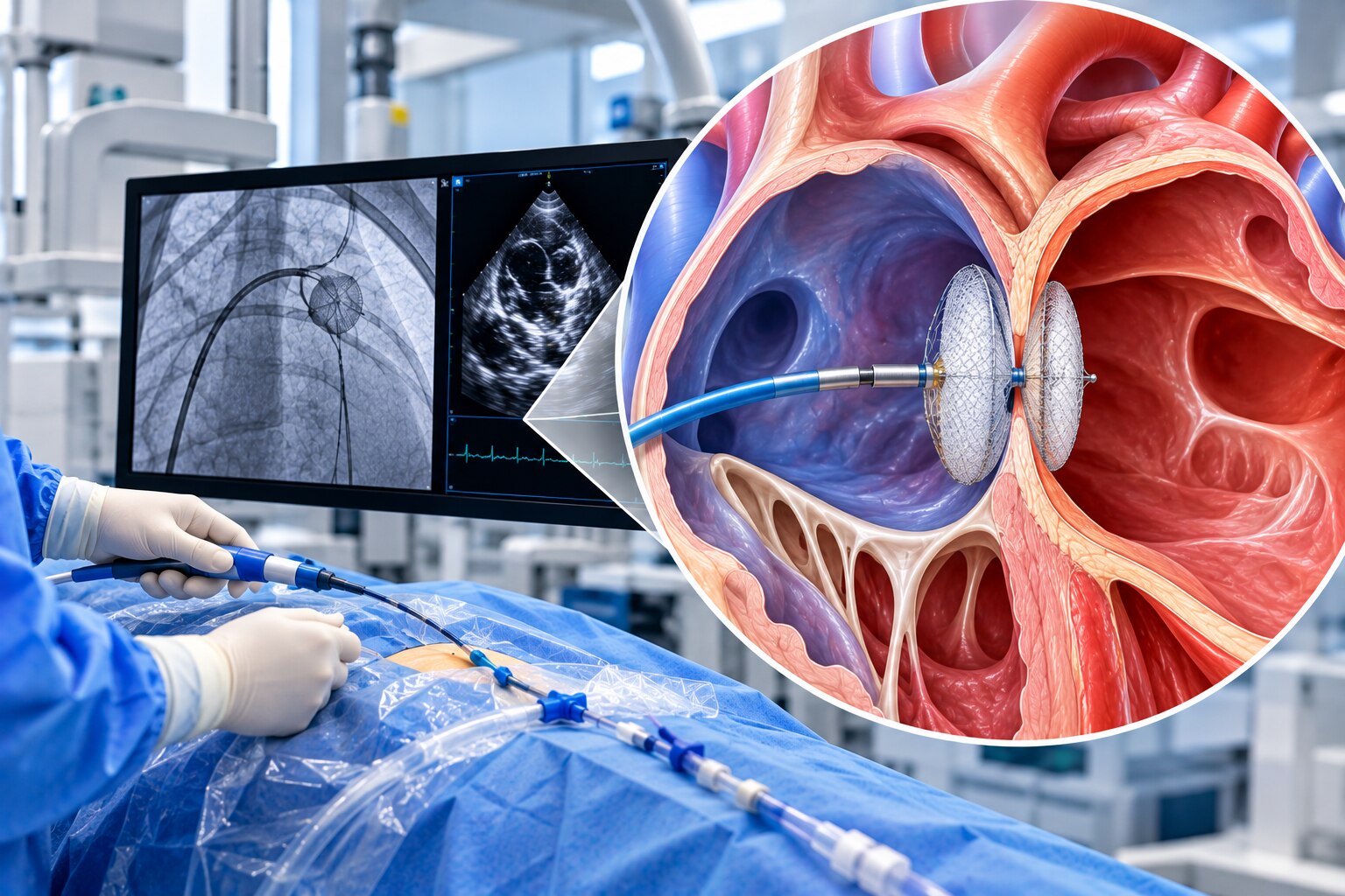
1. Fluoroscopy tracks the catheter through the major veins into the heart, while echocardiographic guidance shows the ASD, septal rims and nearby valves. The catheter crosses the defect from the right atrium into the left atrium.
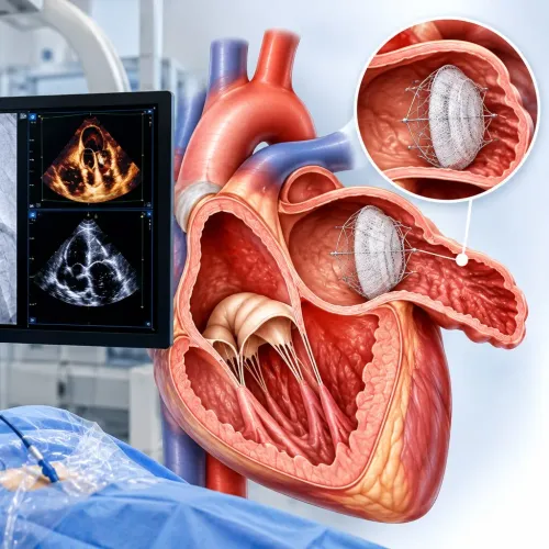
2. The operator advances the delivery system into the left atrium and opens the first disc of the two-disc occluder device. The disc is pulled gently against the left side of the atrial septum; the device’s waist is positioned within the ASD, and the second disc opens on the right-atrial side.
3. Imaging confirms that both discs lie flat and that the device is stable. The team checks for residual blood flow through the defect and ensures that the device does not interfere with the mitral or tricuspid valve, pulmonary veins or other nearby structures. It is released only after these checks pass.
Adults often receive local anaesthetic with conscious or deep sedation. General anaesthesia is used when stillness or transesophageal echocardiography requires it, especially in children. The procedure commonly takes about one to three hours. Because the device travels through a vein, it avoids a chest incision, sternotomy and cardiopulmonary bypass.
What happens to the device and your heart after implantation?
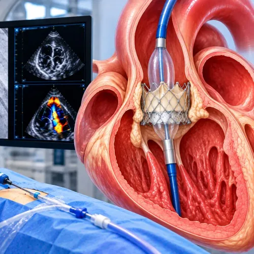
The occluder uses two metal-mesh discs joined by a central waist. The discs rest on opposite sides of the atrial septum, while the waist fills the ASD and blocks the abnormal left-to-right flow immediately after deployment. The opening is not cut out: over weeks to months, endothelial tissue grows across the mesh.
This process, called ASD occluder endothelialization, creates a smoother, incorporated surface.
Most patients remain under overnight observation and leave after recovery from sedation or anaesthesia. Groin bruising, a small amount of soreness, headache, chest discomfort or brief palpitations can occur during recovery.
- Usually settle: mild groin bruising, headache, chest discomfort and transient palpitations.
- Seek urgent care for heavy or expanding groin bleeding, fainting, sudden breathlessness, severe chest pain, sustained arrhythmia, fever, or neurological symptoms. These can signal bleeding, device embolization, erosion, thrombus, infection or a significant rhythm problem.
- Report ongoing breathlessness or a new murmur; these findings can indicate persistent residual shunt or another complication.
A post-procedure echocardiogram and scheduled follow-up scans confirm device position, residual flow and right-heart recovery. The right atrium and ventricle usually reduce their volume load over time. Antiplatelet therapy after ASD closure commonly includes aspirin for about six months when appropriate; do not stop it without medical advice.
Atrial fibrillation or breathlessness can persist if atrial remodeling, lung disease, pulmonary vascular disease or left-heart disease remains.
How should you prepare, and when is specialist assessment essential?
A safe ASD procedure preparation starts with a complete medication review and a clear plan for the day of treatment. Follow the hospital’s fasting before cardiac catheterization instructions exactly; breaking the fast can delay sedation or anaesthesia.
- List every prescription, over-the-counter medicine, vitamin and herbal supplement. Ask whether blood thinners, diabetes medicines or supplements need adjustment.
- Disclose allergies to medicines, latex or iodinated contrast before testing or catheterization.
- Report fever, cough, urinary symptoms, dental infection or any other active infection; the procedure may need postponement.
- Complete requested blood tests, ECG, chest imaging and echocardiography.
- Arrange an adult to take you home and stay with you after sedation or anaesthesia. Do not drive yourself.
A cardiology consultation must confirm the ASD type, measure the septal rims, assess pulmonary pressures and right-heart effects, and identify abnormal valves, veins or other congenital findings. It must also address device closure versus surgery rather than assuming a catheter device is suitable.
| Option | What it means | When it applies |
|---|---|---|
| Device closure | A catheter-delivered occluder seals a suitable defect | Adequate rims, suitable anatomy and acceptable pulmonary resistance |
| Surgical repair | Direct closure through an operation | Large or poorly supported defects, non-secundum anatomy or associated repairs |
In Navi Mumbai, Dr. Rajesh Matta can help coordinate diagnostic testing, congenital-heart assessment, procedural planning and follow-up when anatomy or pulmonary pressures make the decision complex.
Frequently asked questions
Which atrial septal defects can a closure device treat?
A catheter-delivered occluder primarily treats suitable secundum ASDs with enough surrounding tissue to hold the device. Primum, sinus venosus, and coronary sinus defects usually need surgical repair because their anatomy does not provide appropriate device support.
How do doctors decide whether closing the ASD will help?
Doctors assess the defect’s size and location, the septal rims, the left-to-right shunt, right-heart enlargement, pulmonary pressure, symptoms, and other heart conditions using echocardiography and related testing.
How is the occluder guided into the atrial septum?
The cardiologist inserts a delivery sheath through a vein, usually in the groin, and advances it into the heart using X-ray and echocardiographic guidance. The device is opened across the defect, checked for position and valve interference, then released.
What happens to the device and your heart after implantation?
Heart tissue grows over the mesh occluder, securing it in place and closing the abnormal passage. Follow-up echocardiograms check device position, residual flow, rhythm, valves, and right-heart recovery.
How should you prepare, and when is specialist assessment essential?
Ask which medicines to take, when to stop eating and drinking, and whether you need blood tests or dental infection treatment. Seek specialist assessment for breathlessness, palpitations, fainting, reduced exercise tolerance, an abnormal murmur, or an ASD found on imaging.