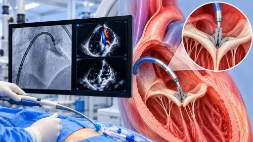
A thickened heart muscle can narrow the route through which blood leaves the left ventricle, causing breathlessness, chest pain, dizziness or fainting during activity. By the end, you will understand how alcohol septal ablation reduces that obstruction, which tests establish whether it is responsible for your symptoms, and how specialists choose it compared with surgical myectomy.
Key takeaways
- A thickened septum can restrict blood leaving the left ventricle.
- Echocardiography measures whether the LVOT gradient explains your symptoms.
- Alcohol ablation reduces septal muscle through a targeted catheter procedure.
- Compare eligibility, risks, and recovery with surgical myectomy before deciding.
How a Thickened Septum Blocks Blood Flow
In obstructive hypertrophic cardiomyopathy, a thickened ventricular septum narrows the left-ventricular outflow tract (LVOT), the passage through which blood leaves the left ventricle for the aorta. The obstruction is dynamic: it worsens as the heart contracts, especially during exercise, when the ventricle beats faster and has less time to fill.
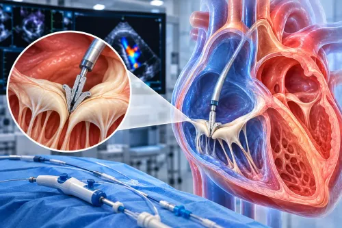
The contracting blood flow can pull the front leaflet of the mitral valve toward the septum, a movement called systolic anterior motion of the mitral valve. The leaflet can further narrow the LVOT and reduce forward blood flow.
It can also stop the mitral valve from closing properly, allowing blood to leak backward as mitral regurgitation.
This combination raises pressure inside the heart and limits how much blood reaches the body. Symptoms can include:
- Breathlessness or reduced exercise tolerance because blood output cannot rise normally during exertion.
- Chest pain from the increased workload of the thickened heart muscle.
- Dizziness or fainting when blood flow falls sharply, particularly during exertion or dehydration.
- Palpitations or worsening breathlessness when mitral regurgitation adds to the pressure burden.
A visible septal bulge or measured gradient alone does not prove it causes symptoms. Doctors must match the obstruction to your breathlessness, chest discomfort, dizziness, or exercise limitation before considering septal reduction. Alcohol septal ablation targets the obstructing basal muscle, allowing the LVOT to widen over time and often reducing associated mitral leakage.
How Doctors Prove the Gradient Is Causing Your Symptoms
A clinically important obstruction requires two findings: a peak LVOT gradient of at least 50 mmHg at rest or with physiologic provocation, and symptoms that match the obstruction. A gradient alone does not justify septal reduction.
| Test | What doctors measure | What the result means |
|---|---|---|
| Transthoracic echocardiography at rest | Doppler blood-flow speed, resting gradient, septal thickness, systolic anterior motion and mitral regurgitation | Confirms obstruction present without exertion |
| Valsalva maneuver during echocardiography | The provoked gradient while you strain and reduce venous return | Exposes dynamic obstruction hidden by a low resting gradient |
| Exercise echocardiography | The LVOT gradient, blood pressure and mitral regurgitation during exercise | Shows whether obstruction rises when breathlessness or chest discomfort occurs |
| Cardiac catheterisation | Pressure difference across the LVOT when non-invasive results remain uncertain | Provides an invasive confirmation before treatment |
A normal or modest resting gradient does not exclude clinically important obstruction. If symptoms suggest obstructive hypertrophic cardiomyopathy and the resting gradient is below 50 mmHg, doctors use Valsalva and, when appropriate, exercise echocardiography rather than relying on the resting scan.
The team then checks whether obstruction explains your limitation. Breathlessness that occurs with a marked exercise-induced gradient supports treatment; breathlessness from lung disease, atrial fibrillation, diastolic dysfunction or deconditioning does not. Treating an unrelated gradient exposes you to procedural risk without addressing the real problem.
What Alcohol Septal Ablation Does Inside the Heart
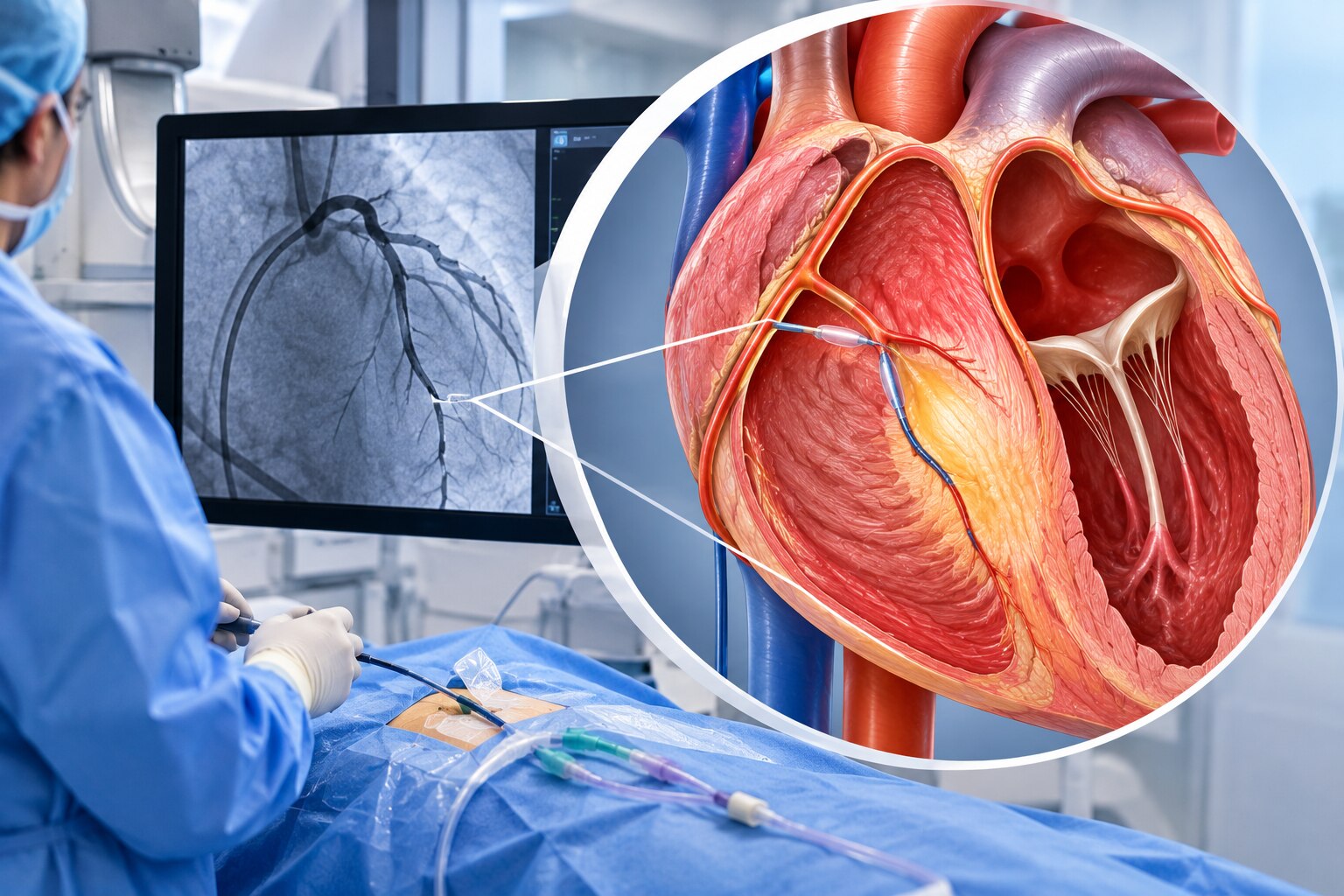
A catheter is passed through an artery in the wrist or groin into the coronary arteries. The operator identifies the septal perforator artery feeding the obstructing basal septum, inflates a temporary balloon to isolate it, and injects contrast to confirm that the ballooned branch supplies the correct muscle.
After that confirmation, a measured amount of medical alcohol is delivered through the catheter. The alcohol creates a controlled infarction, intentionally injuring a small area rather than blocking a normal coronary artery. That injured muscle later becomes septal scar tissue, reducing the septum’s bulk and improving blood flow through the left-ventricular outflow tract.
The pressure gradient may fall during or soon after the procedure, but the full benefit is not immediate. Scar formation and remodelling continue over weeks to months, so breathlessness and exercise capacity often improve gradually. Follow-up echocardiography checks the gradient and heart function.
ASA is a deliberate myocardial-infarction procedure, not a risk-free catheter treatment. Risks include:
- Injury or spasm of the coronary artery
- Alcohol spreading beyond the intended septum
- Temporary or permanent heart block requiring a pacemaker
- Ventricular arrhythmias
- Ventricular septal defect
- Bleeding or vascular damage at the catheter site
- Stroke or death
An experienced hypertrophic cardiomyopathy team reduces these dangers through precise imaging, monitoring and immediate access to electrophysiology, surgery and intensive care.
Who Is a Suitable Candidate—and Who Is Not
Alcohol septal ablation fits an adult with symptomatic obstructive HCM despite appropriate medical treatment and a left-ventricular outflow-tract gradient of at least 50 mmHg at rest or with physiologic provocation. A high gradient without troublesome symptoms is not, by itself, a reason for septal reduction.
The anatomy must point to a treatable basal septal target. Coronary imaging should show a septal perforator branch that reliably supplies the muscle causing obstruction; the operator temporarily blocks that branch and confirms its territory before injecting alcohol. Reducing this muscle can lessen systolic anterior motion and obstruction-related mitral regurgitation.
ASA is a poor fit when the supplying branch is very small, cannot be safely identified, or does not reach the obstructing segment. Inadequate septal thickness also leaves too little target tissue for a controlled result. Surgical myectomy may be more appropriate when:
- The obstruction pattern is not confined to a reachable basal septal segment.
- Severe mitral regurgitation arises from intrinsic valve disease rather than systolic anterior motion.
- Another cardiac operation is already needed.
- The coronary anatomy makes alcohol delivery unsafe or unreliable.
Choose the procedure through an experienced HCM team with advanced imaging, electrophysiology, surgery and intensive-care support. Anatomy, symptom burden and your treatment priorities should drive the decision—not a preference for a catheter procedure alone.
Choosing Between Ablation and Surgical Myectomy
The choice between alcohol septal ablation versus surgical myectomy depends on your septal anatomy, coronary blood supply, other heart problems, and tolerance for each procedure—not on which treatment sounds newer. Make the decision with a multidisciplinary HCM team that performs septal reduction regularly and has advanced imaging, electrophysiology, surgery, and intensive-care support.
| Option | Main advantage | When it fits |
|---|---|---|
| Alcohol septal ablation | Catheter-based treatment that creates a controlled scar in the obstructing septum | A suitable septal artery reliably supplies the target, and you do not need another cardiac operation |
| Surgical myectomy | Directly removes excess septal muscle and can correct associated valve or structural problems | The septal artery is too small or unsuitable, obstruction is complex, or another operation is needed |
| Key trade-off | Both can lower the LVOT gradient and improve exertional symptoms; neither removes the inherited HCM substrate | Choose after reviewing imaging, symptoms, risks, and your preferences |
Alcohol septal ablation carries a distinctive risk of new conduction disease. Complete heart block can require a permanent pacemaker, particularly when bundle-branch disease already exists. Myectomy has surgical risks, including bleeding, infection, stroke, and a longer recovery.
Neither procedure guarantees that breathlessness or fatigue will disappear. Diastolic dysfunction, atrial fibrillation, small-vessel ischemia, lung disease, or deconditioning can continue causing symptoms.
After either treatment, HCM follow-up includes ECGs, symptom review, rhythm monitoring when indicated, and echocardiography to reassess the gradient and mitral regurgitation. Dr. Rajesh Matta can help coordinate that longer-term review, including medication decisions and referral for device or rhythm care when findings require it.
Frequently asked questions
How does a thickened septum block blood flow?
It narrows the left-ventricular outflow tract, creating a dynamic obstruction that can worsen during exercise.
How do doctors prove the LVOT gradient causes symptoms?
Doctors use cardiac imaging and exercise assessment to measure the gradient and relate it to symptoms such as breathlessness, chest pain, or fainting.
What does alcohol septal ablation do inside the heart?
A catheter delivers alcohol into a selected septal artery, reducing the targeted heart muscle and widening the outflow tract.
Who is a suitable candidate for alcohol septal ablation?
It suits selected adults with symptomatic obstructive hypertrophic cardiomyopathy despite medication and suitable septal artery anatomy.
How does alcohol septal ablation compare with surgical myectomy?
Both treatments reduce LVOT obstruction; the choice depends on anatomy, valve findings, comorbidities, surgical risk, and local expertise.