A coronary narrowing that looks severe on an angiogram does not always restrict blood flow enough to justify intervention, while a moderate-looking lesion can still cause ischemia. By the end, you will understand how FFR measures pressure loss, how to interpret the result, what the procedure feels like, and when the finding should or should not change treatment.
Key takeaways
- FFR compares pressure beyond a narrowing with pressure before it during maximum blood flow.
- An FFR of 0.80 or less often supports treatment of a significant narrowing.
- Angiograms show artery shape but cannot reliably measure the narrowing’s effect on blood flow.
- Ask whether FFR, imaging, stress testing, or another test best answers your clinical question.
What FFR measures and how the pressure ratio is calculated
Fractional flow reserve measures how much a coronary narrowing reduces pressure available to the heart muscle during maximum blood flow. Its FFR meaning is functional, not visual: it estimates the artery’s maximum flow capacity with the narrowing compared with flow if that obstruction were absent.
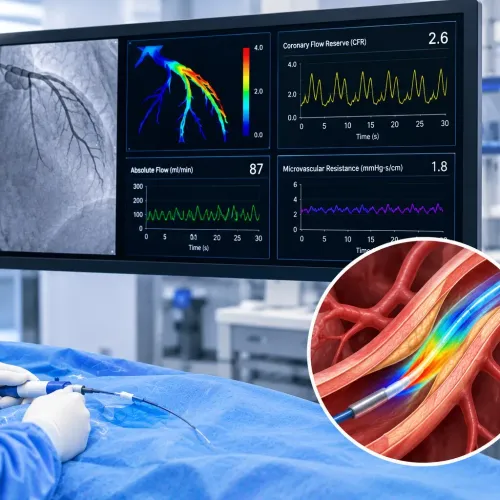
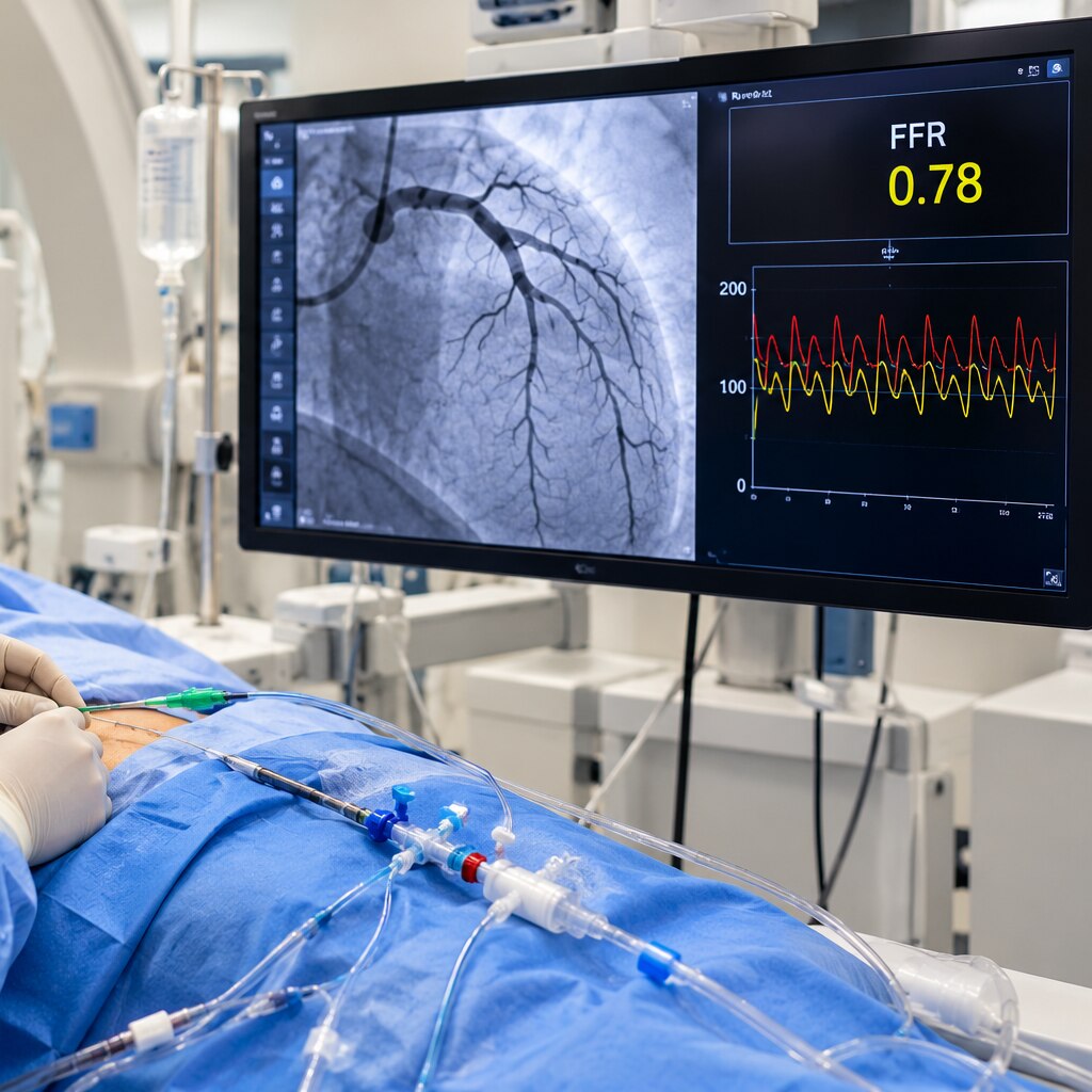
During catheterisation, the clinician advances a pressure-sensing guidewire beyond the narrowing. With the sensor at the guide-catheter tip, pressures are equalized, then the wire records distal coronary pressure while the catheter records aortic pressure. Medication induces maximal hyperemia, when the coronary vessels are dilated and the narrowing’s effect becomes measurable.
The calculation is distal coronary pressure Pd/Pa: FFR equals Pd divided by Pa. Pd is the pressure beyond the lesion; Pa is the aortic pressure at the guide catheter. The pressure difference across coronary narrowing appears as the fall from Pa to Pd, but the ratio is the reported result.
Before accepting the number, the operator checks for pressure-wire drift by returning the sensor to the guide-catheter tip and confirming that the pressures still match. Inadequate hyperemia or guide-catheter damping can distort the measurement. FFR does not come from an angiogram alone; the pressure wire and hyperemia are essential.
How to interpret 0.80—and why an angiogram can mislead
An FFR of 0.80 is a decision boundary, not a biological cliff. A value at or below the FFR 0.80 threshold indicates that the narrowing is generally a hemodynamically significant stenosis and is more likely to benefit from revascularisation.
A value above 0.80 often supports deferring treatment when symptoms, test results and the clinical situation agree.
- FFR ≤0.80: Treat the lesion as potentially flow-limiting and discuss revascularisation alongside the patient’s symptoms and other findings.
- FFR >0.80: Defer stenting when the measurement is technically sound and no other evidence indicates ischaemia from that lesion.
- FFR close to 0.80: Do not treat the number as an automatic yes-or-no instruction. Check pressure-wire drift, hyperaemia, guide-catheter damping and the wider clinical picture.
The key angiography versus FFR difference is that angiography shows the artery’s outline, while FFR measures the narrowing’s effect on pressure and flow. A severe-looking coronary narrowing can preserve enough blood flow to produce an FFR above 0.80, especially when the lesion is short or the reference artery is difficult to judge.
A visually moderate narrowing can produce an FFR at or below 0.80 when plaque length, vessel geometry or additional disease increases resistance.
Tachycardia, anaemia, low blood pressure, acute coronary syndrome and a recent myocardial infarction can also distort interpretation. FFR assesses one lesion, not overall plaque risk, microvascular disease or vasospastic angina.
What happens during FFR, from preparation to recovery
The FFR procedure steps usually take place during coronary catheterisation. You will receive consent, monitoring, local anaesthetic, and contrast dye; tell the team about allergies, kidney disease, bleeding problems, and medicines such as anticoagulants.
1. The cardiologist places a catheter through a radial or femoral access site and positions it at the coronary artery opening. The pressure-sensing guidewire passes across the narrowing. With its sensor at the guide-catheter tip, the team equalises the wire and catheter pressures.
2. The wire records pressure beyond the narrowing while the catheter records aortic pressure. The team gives adenosine by intravenous infusion or directly into the coronary artery to produce maximal blood flow, then calculates the pressure ratio.
3. Adenosine effects can include a brief warm flush, chest pressure, breathlessness, headache, or an awareness of a slow heartbeat. Transient atrioventricular block can occur; these effects usually stop quickly when the medicine ends.
4. The operator checks the tracing for pressure-wire drift and repeats measurements if needed. Inadequate hyperaemia can produce a falsely high result, so the team may adjust the infusion or reassess the recording.
Afterward, the catheter and wire are removed. Pressure or a closure device controls bleeding at the access site, and staff monitor your pulse, blood pressure, and puncture area before discharge. Expect soreness or bruising; seek urgent help for persistent bleeding, a growing swelling, a cold or numb limb, severe chest pain, or breathlessness.
Rare risks include artery damage, irregular rhythm, allergic reaction to contrast, kidney injury, heart attack, or stroke.
When FFR is useful—and when another test gives better information
FFR is most useful for an angiographically intermediate narrowing when ischemia has not already been established. iFR versus FFR offers two accepted invasive options: iFR needs no drug-induced hyperemia, while FFR measures during maximal hyperemia. Revascularization is generally considered at FFR ≤0.80 or iFR ≤0.89.
| Option | Best information | Limitation |
|---|---|---|
| iFR | Pressure-based, lesion-specific significance without hyperemia | Does not show plaque structure |
| IVUS and OCT | Vessel diameter, plaque burden, calcium, dissection and stent detail | Anatomy does not prove that blood flow is restricted |
| Stress testing | Whether exercise or medication provokes ischemia in the heart | Usually less precise about which coronary lesion is responsible |
| Coronary CT angiography and FFR-CT | Noninvasive anatomy; FFR-CT estimates lesion-specific pressure effects from CT images | Image quality, heavy calcium, motion and complex disease can reduce reliability |
FFR is harder to interpret with serial or diffuse narrowings, left-main or ostial disease, severe calcification, and a recently infarcted or acutely unstable artery. Microvascular disease, tachycardia, anaemia, hypotension and inadequate hyperemia can reduce coronary flow and make a significant narrowing appear less abnormal.
Check for pressure-wire drift, guide-catheter damping and a stable hyperemic response before accepting a borderline result. Compare it with symptoms, ECG, stress testing and imaging. A normal FFR does not exclude vasospasm, microvascular angina, plaque elsewhere or future cardiovascular risk.
How the result should change a treatment decision
An FFR of 0.80 or less supports revascularisation when symptoms, anatomy and procedural risk fit, but it does not make PCI automatic. In stable coronary disease, FFR-guided PCI can reduce urgent revascularisation compared with medical therapy alone; it has not guaranteed fewer deaths or heart attacks.
An FFR above 0.80 usually supports deferring PCI and continuing guideline-directed medical therapy. That means no change to the blockage itself when symptoms are controlled and no other finding demands intervention. The threshold is a decision boundary, not a biological cliff.
Choose further evaluation when the value is close to 0.80, symptoms and the result disagree, or technical quality is uncertain. Check the pressure-wire trace for drift, confirm adequate hyperaemia, and consider iFR, intravascular imaging, stress testing or another assessment.
Acute coronary syndrome, recent infarction, anaemia, hypotension and severe microvascular dysfunction can distort interpretation; review FFR contraindications and limitations before relying on the number. Dr. Rajesh Matta can help place the result alongside your symptoms, ECG, imaging and overall treatment risk rather than treating it as an isolated yes-or-no answer.
- What decision will this measurement change if the result is above or below 0.80?
- Is iFR or another test safer or more informative for my anatomy?
- How will you check for pressure-wire drift and adequate hyperaemia?
- If PCI is advised, what benefit, stent-related treatment and alternatives should I expect?
Related service
Coronary Angiography & Angioplasty Coronary Angiography & Angioplasty At Anahat Advanced and Comprehensive Cardiac Clinic, Dr. Rajesh Matta provides advanced care for heart blockages through Coronary Angiography and... View service → |
Frequently asked questions
What does fractional flow reserve measure?
FFR measures how much a coronary artery narrowing reduces pressure available to the heart muscle during maximum blood flow. It compares pressure beyond the narrowing with pressure before it.
What does an FFR of 0.80 mean?
An FFR of 0.80 indicates that pressure beyond the narrowing is 80% of the pressure before it during maximum blood flow. Values at or below 0.80 often support treating the narrowing, alongside symptoms and other findings.
Why can an angiogram mislead doctors about a coronary narrowing?
An angiogram shows the artery’s outline but does not directly measure how much the narrowing limits blood flow. A visually severe narrowing can have limited functional impact, while a less dramatic narrowing can reduce flow significantly.
What happens during an FFR procedure?
After preparation, a catheter is guided to the coronary artery. A pressure wire measures pressure before and beyond the narrowing during maximum blood flow, after which the equipment is removed and you are monitored during recovery.
When is another test better than FFR?
Another test may provide better information when the question concerns plaque structure, blood-flow patterns, exercise-related symptoms, or whether the artery is suitable for FFR measurement. Options include intravascular imaging, stress testing, and other physiological assessments.